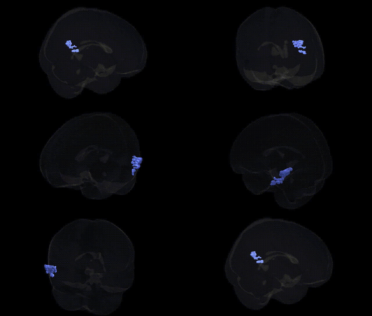
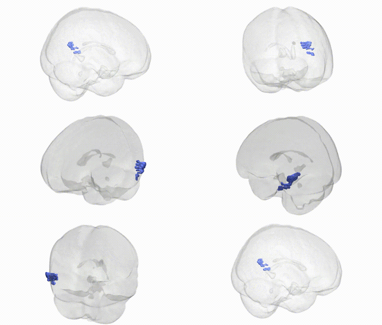
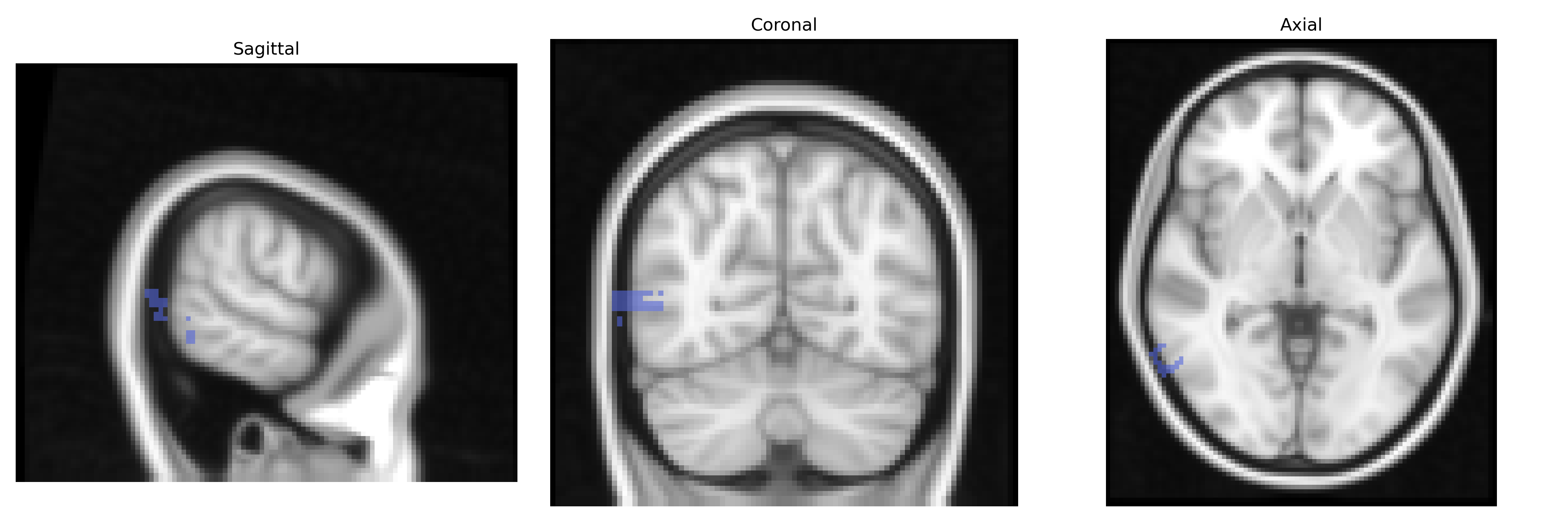
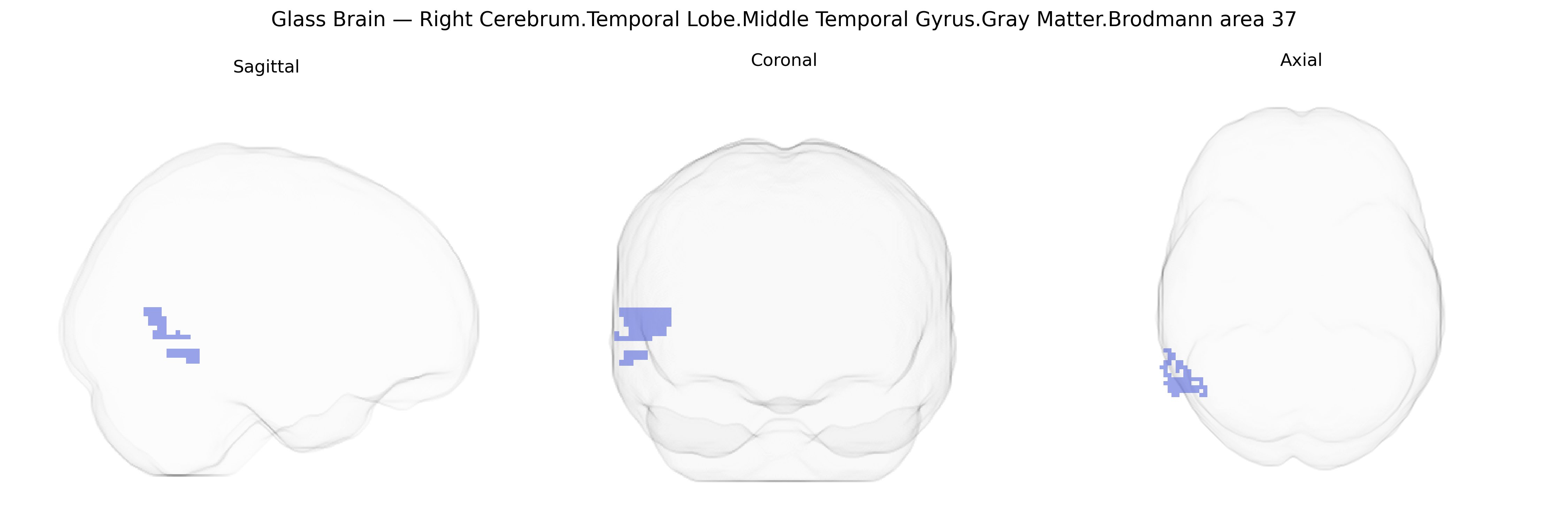
Bilateral Right Cerebrum.Temporal Lobe.Middle Temporal Gyrus.Gray Matter.Brodmann area 37 corresponds to a cytoarchitectonic and functional territory situated in the posterior portion of the temporal lobe, extending over the fusiform and adjacent inferior/middle temporal regions. Brodmann area 37 is implicated in high-level visual processing, particularly the recognition and categorization of complex visual stimuli such as faces, objects, and written words, and contributes to multimodal integration linking visual inputs with semantic memory and language-related processes. Neuronal populations in this region exhibit specialization for category-selective processing (e.g., faces and words), and the area maintains dense reciprocal connections with occipital visual cortices, ventral temporal regions, and language networks, supporting its role at the interface of perception and meaning. There is no direct link for this full Talairach label; a related structure is Brodmann area 37.
The bilateral right middle temporal gyrus gray matter in Brodmann area 37 (posterior inferior/middle temporal cortex, overlapping the fusiform gyrus and visual word/form and face-processing regions) has been implicated in several genetic and GWAS-based associations, largely via imaging‑genetics and disorder‑focused studies rather than region‑specific genome-wide screens. Variants in genes linked to synaptic function and neurodevelopment—such as BDNF (e.g., Val66Met), COMT, and DISC1—have been repeatedly associated with structural and functional differences in temporal and fusiform regions that encompass BA37, with downstream effects on language, semantic processing, and visual recognition. Large ENIGMA and UK Biobank imaging GWAS have identified multiple common variants (including loci near genes such as KIAA0586, WNT3, and others) associated with temporal lobe gray matter volume and cortical thickness, which likely include BA37 but do not isolate this subregion. BA37‑adjacent fusiform cortex shows genetic modulation in face‑selective activation and social cognition, including associations with autism‑related genes (e.g., CNTNAP2, SHANK3), consistent with findings that temporal/fusiform morphology and activity are altered in autism spectrum disorder, schizophrenia, and dyslexia, each of which has polygenic architectures involving synaptic, neuronal migration, and transcriptional regulation pathways. In Alzheimer’s disease and other dementias, GWAS‑identified risk loci such as APOE, CLU, and PICALM are associated with atrophy and hypometabolism in temporal association cortices, including BA37, parallel to deficits in object and word recognition. Overall, genetic influences on BA37 appear to be largely polygenic and shared with broader temporal and fusiform networks, contributing to variability in language, reading, face processing, social cognition, and susceptibility to neurodevelopmental and neurodegenerative disorders.
Overview generated by GPT-4o (2026).
Region ID: 328
Hemisphere: bilateral
Atlas: Talairach labels 2mm
Full Quality Version: Download MP4
Full Quality Version: Download MP4
Wali Sidiqyar*, Gaurav Rudravaram*, Elyssa M. McMaster, Trent M. Schwartz, Adam M. Saunders, Kurt G. Schilling, Bennett A. Landman "Introducing SPINS: A Shared Public Visualization Library of Neuroanatomical Structures." Medical Imaging with Deep Learning- short paper
This resource is licensed under CC0 1.0 Universal (Public Domain).