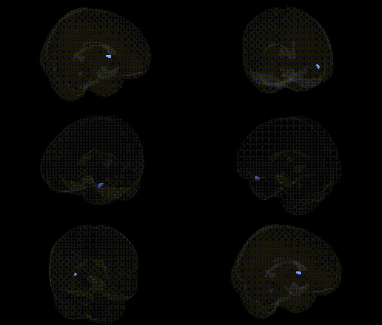
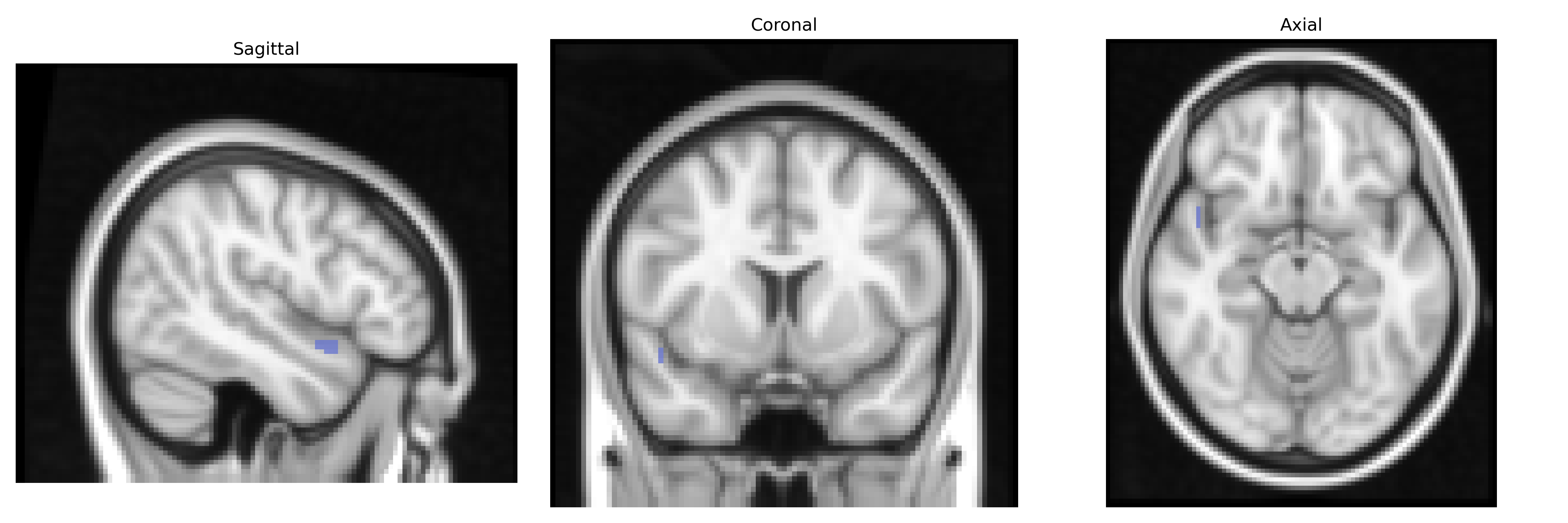
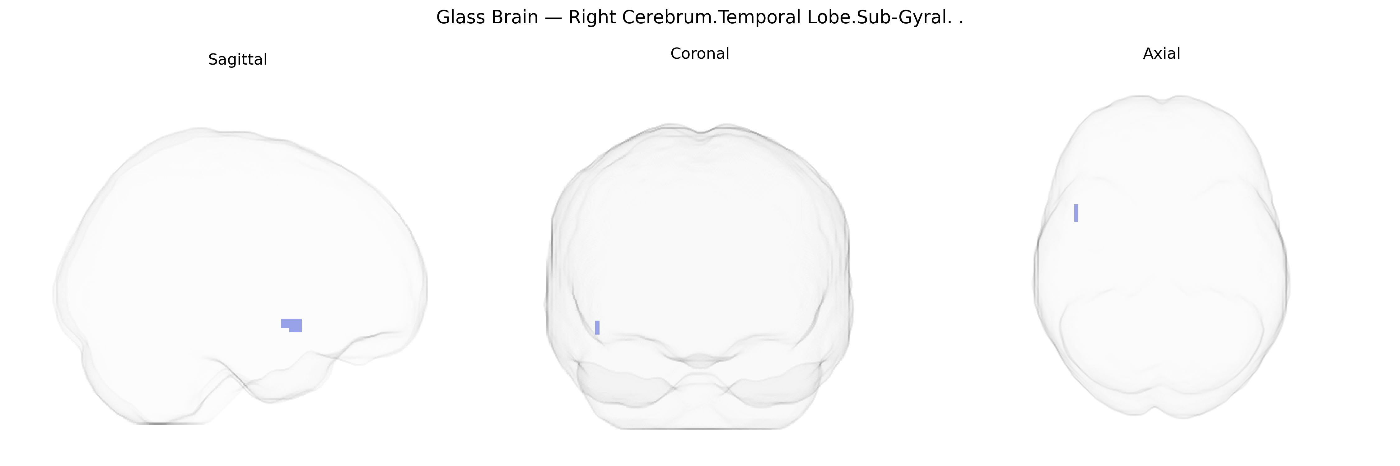
The bilateral Right Cerebrum.Temporal Lobe.Sub-Gyral region in the Talairach 2 mm atlas refers to subcortical white matter and deep cortical tissue located beneath the gyri of the right temporal lobe, including fibers interconnecting temporal lobe cortices with other association areas and limbic structures. Functionally, this area supports transmission and integration of auditory, language, and memory-related information by linking superior, middle, and inferior temporal gyri with hippocampal and frontal regions, and is involved in higher-order processing of complex sounds, semantic content, and aspects of visual object recognition. As a white matter–rich sub-gyral zone rather than a named cortical area, it does not have a specific Wikipedia article; a closely related structure is the Temporal lobe.
Genetic associations involving the bilateral right temporal sub-gyral region (as defined in the Talairach 2 mm atlas) arise primarily from imaging genetics and GWAS of brain structure and function, though most studies target broader temporal lobe or superior/middle/inferior temporal gyri rather than the sub-gyral label specifically. Variants in genes related to synaptic function, neurodevelopment, and axonal guidance (e.g., BDNF, DISC1, CNTNAP2, NRG1) have been repeatedly implicated in temporal lobe volume and connectivity, and in susceptibility to temporal lobe epilepsy, schizophrenia, and autism spectrum disorders, which often show structural and functional abnormalities in temporal subcortical and sub-gyral areas. Large-scale GWAS of cortical thickness and surface area (such as ENIGMA and UK Biobank efforts) have identified multiple loci—frequently near genes like MEF2C, TBR1, and various cadherins and protocadherins—associated with temporal lobe morphometry, some of which overlap with risk loci for cognitive performance, educational attainment, and language-related phenotypes. Temporal sub-gyral tissue also contributes to networks underlying auditory and language processing, social cognition, and memory; thus, genetic variants influencing these traits and related disorders (e.g., primary progressive aphasia, frontotemporal dementia, and Alzheimer’s disease via genes such as APOE and CLU) are indirectly linked to this region through convergent evidence of altered temporal lobe structure and metabolism. Overall, while few studies isolate the “sub-gyral” compartment at 2 mm Talairach resolution, converging GWAS and candidate-gene findings indicate that polygenic influences on neurodevelopment, synaptic plasticity, and neurodegeneration contribute to variability and disease-related changes in this bilateral right temporal sub-gyral region.
Overview generated by GPT-4o (2026).
Region ID: 384
Hemisphere: bilateral
Atlas: Talairach labels 2mm
Full Quality Version: Download MP4
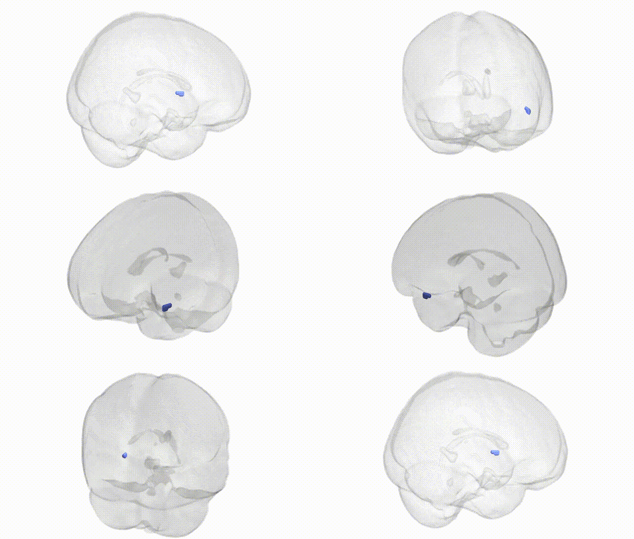
Full Quality Version: Download MP4
Wali Sidiqyar*, Gaurav Rudravaram*, Elyssa M. McMaster, Trent M. Schwartz, Adam M. Saunders, Kurt G. Schilling, Bennett A. Landman "Introducing SPINS: A Shared Public Visualization Library of Neuroanatomical Structures." Medical Imaging with Deep Learning- short paper
This resource is licensed under CC0 1.0 Universal (Public Domain).