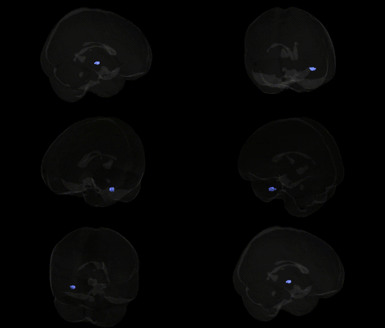
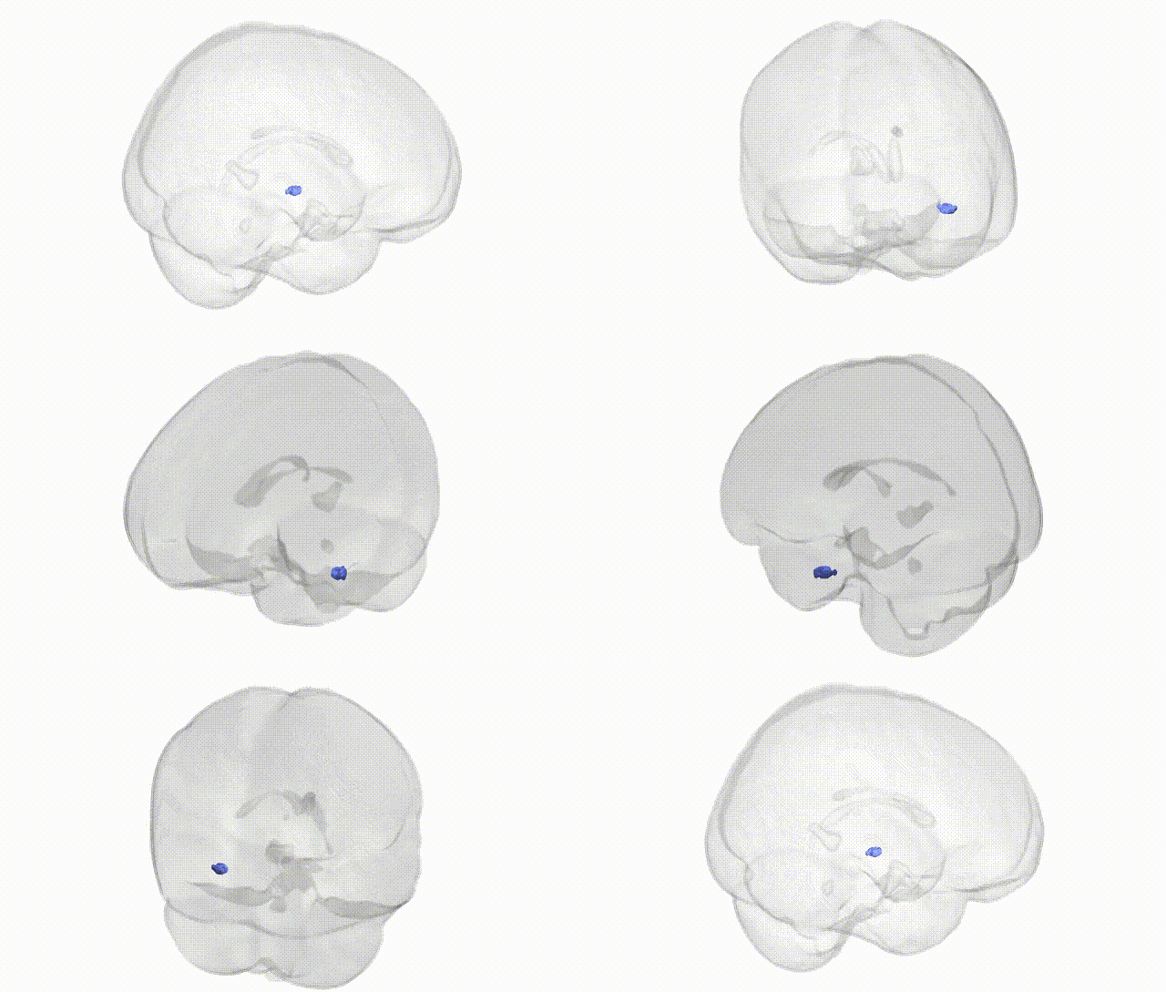
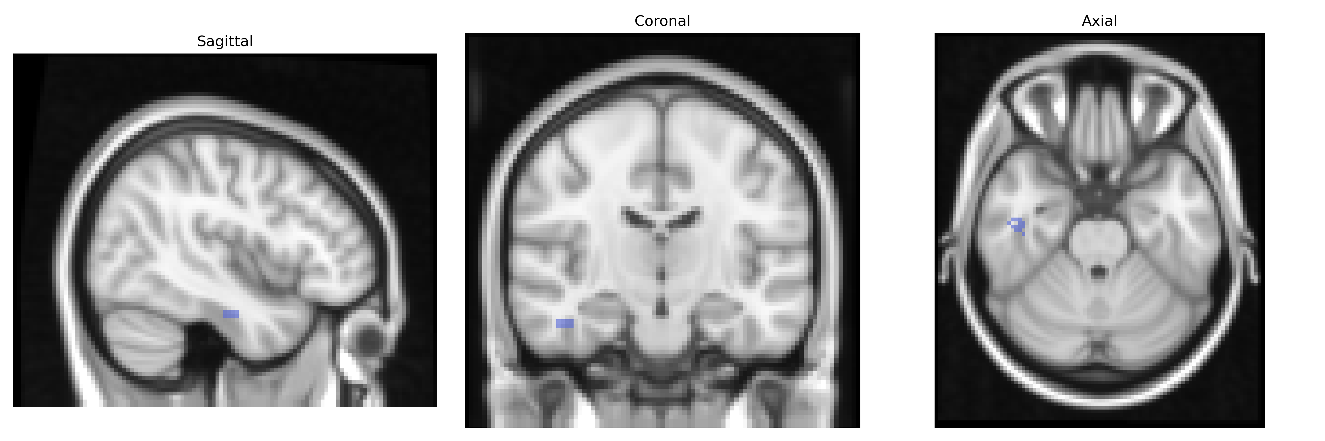
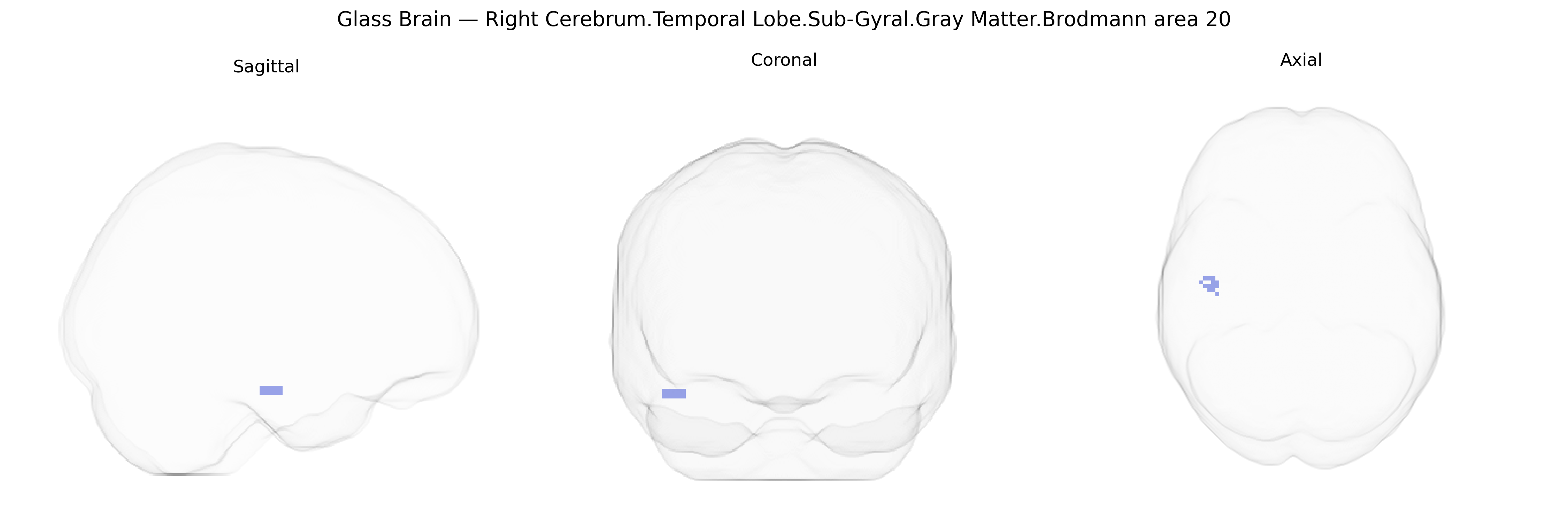
The bilateral Right Cerebrum.Temporal Lobe.Sub-Gyral.Gray Matter.Brodmann area 20 corresponds to ventral temporal association cortex located in the inferior temporal gyrus, primarily involved in high-level visual processing and object recognition. Cytoarchitectonically defined by Brodmann, area 20 contains densely packed pyramidal neurons and forms part of the ventral visual stream (“what” pathway), contributing to the integration of complex visual features into coherent percepts and supporting semantic memory and language-related processes through connections with adjacent temporal and limbic regions. Functionally, it participates in recognition of faces, scenes, and other complex stimuli, and lesions in this region have been associated with deficits in visual agnosia and disturbances of associative visual memory. There is no direct Wikipedia article for Brodmann area 20; see the related structure Inferior temporal gyrus.
The bilateral right temporal lobe sub-gyral gray matter corresponding to Brodmann area 20 (inferior temporal cortex involved in high-level visual and semantic processing) has been implicated in multiple genetic and GWAS-derived associations through its structure, function, and connectivity rather than via variants explicitly tied to the Talairach atlas label. Large neuroimaging GWAS (e.g., ENIGMA, UK Biobank) have identified common variants in genes related to neuronal development, synaptic function, and axonal guidance—such as BDNF, HMGA2, MAPT, and multiple loci near microtubule and cell-adhesion genes—that influence temporal lobe cortical thickness and surface area, with downstream effects on BA20-like regions. Structural and functional variation in inferior temporal cortex has been repeatedly associated with genetic risk for neuropsychiatric and neurodevelopmental disorders, including schizophrenia (e.g., risk loci in the MHC region and synaptic genes affecting temporal lobe volume and activation), autism spectrum disorder (rare CNVs and common variants impacting temporal and ventral visual pathways), major depressive disorder (variants in neurotrophic and stress-response genes influencing temporal cortex morphology), and Alzheimer’s disease (APOE ε4 and other AD risk loci associated with temporal atrophy involving ventral temporal areas). GWAS of cognitive traits such as general intelligence, reading ability, and face/object recognition performance also show polygenic influences on ventral temporal regions, where BA20 contributes to semantic and visual object processing. While no single variant is uniquely specific to the “right cerebrum temporal lobe sub-gyral BA20” Talairach parcel, convergent genetic imaging and disorder GWAS evidence supports this region as a downstream target of polygenic risk architectures affecting temporal lobe structure, higher-order visual recognition, language-related semantic processes, and vulnerability to psychiatric and neurodegenerative disease.
Overview generated by GPT-4o (2026).
Region ID: 172
Hemisphere: bilateral
Atlas: Talairach labels 2mm
Full Quality Version: Download MP4
Full Quality Version: Download MP4
Wali Sidiqyar*, Gaurav Rudravaram*, Elyssa M. McMaster, Trent M. Schwartz, Adam M. Saunders, Kurt G. Schilling, Bennett A. Landman "Introducing SPINS: A Shared Public Visualization Library of Neuroanatomical Structures." Medical Imaging with Deep Learning- short paper
This resource is licensed under CC0 1.0 Universal (Public Domain).