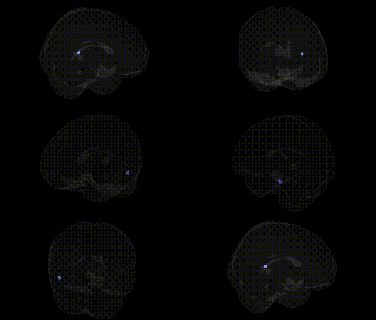
The bilateral Right Cerebrum.Temporal Lobe.Sub-Gyral.Gray Matter.Brodmann area 37 corresponds to a portion of the fusiform and adjacent inferior temporal cortex implicated in high-level visual and multimodal processing. Brodmann area 37 is strongly associated with visual object recognition, face and word-form processing, and the integration of visual information with semantic memory, often considered part of the ventral “what” visual stream. Functionally, this region contributes to the identification and categorization of complex stimuli, including faces and written language, and plays a role in linking perceptual input to stored knowledge. Clinically, damage to area 37 can lead to deficits such as prosopagnosia (impaired face recognition) or visual agnosias. There is no direct link for this exact Talairach label; a closely related and encompassing structure is Fusiform gyrus.
The bilateral right cerebrum temporal lobe sub-gyral gray matter corresponding to Brodmann area 37 (inferior temporal/fusiform region) has been implicated in several genetic and GWAS-based findings, largely through imaging genetics and disorder-focused studies rather than region-specific genome scans. BA37 is central to visual word and object recognition and faces within the fusiform gyrus, and structural and functional variation in this area has been associated with common psychiatric and neurodevelopmental disorders including autism spectrum disorder (ASD), schizophrenia, and major depressive disorder, often in relation to polygenic risk scores and risk loci such as CACNA1C, MIR137, and others that modulate temporal lobe volume and connectivity. Imaging GWAS of cortical thickness and surface area have identified heritable variation in inferior temporal/fusiform regions linked to loci near genes involved in neurodevelopment and synaptic function (for example, variants near PAX6, KIAA0586, and other developmental regulators), although results are typically reported at the level of the inferior temporal or fusiform cortex rather than Talairach-specific BA37 subregions. Rare variant and familial studies have connected mutations in genes critical for cortical development (e.g., TBR1, DCC, RELN) with temporal lobe malformations and atypical cytoarchitecture overlapping BA37, while dyslexia and language-related GWAS and imaging genetics studies show that alleles in genes such as DCDC2, KIAA0319, and FOXP2 influence activation and structure in left-lateralized BA37 homologs, with some evidence of bilateral fusiform involvement. Alzheimer’s disease and frontotemporal dementia genetics (including APOE ε4 and MAPT haplotypes) have been related to patterns of atrophy that frequently involve ventral temporal cortex, including BA37, and GWAS of face recognition and social cognition traits have shown heritable fusiform activation patterns linked to polygenic architectures affecting synaptic plasticity genes, though individual loci remain incompletely resolved. Overall, genetic associations for this specific Talairach-defined BA37 region are indirect, emerging from broader fusiform/inferior temporal lobe imaging genetics in psychiatric, cognitive, and degenerative disorders, with convergent evidence that common and rare variants influencing neurodevelopment, synaptic signaling, and cortical morphology contribute to structural and functional variability in this area.
Overview generated by GPT-4o (2026).
Region ID: 333
Hemisphere: bilateral
Atlas: Talairach labels 2mm
Full Quality Version: Download MP4
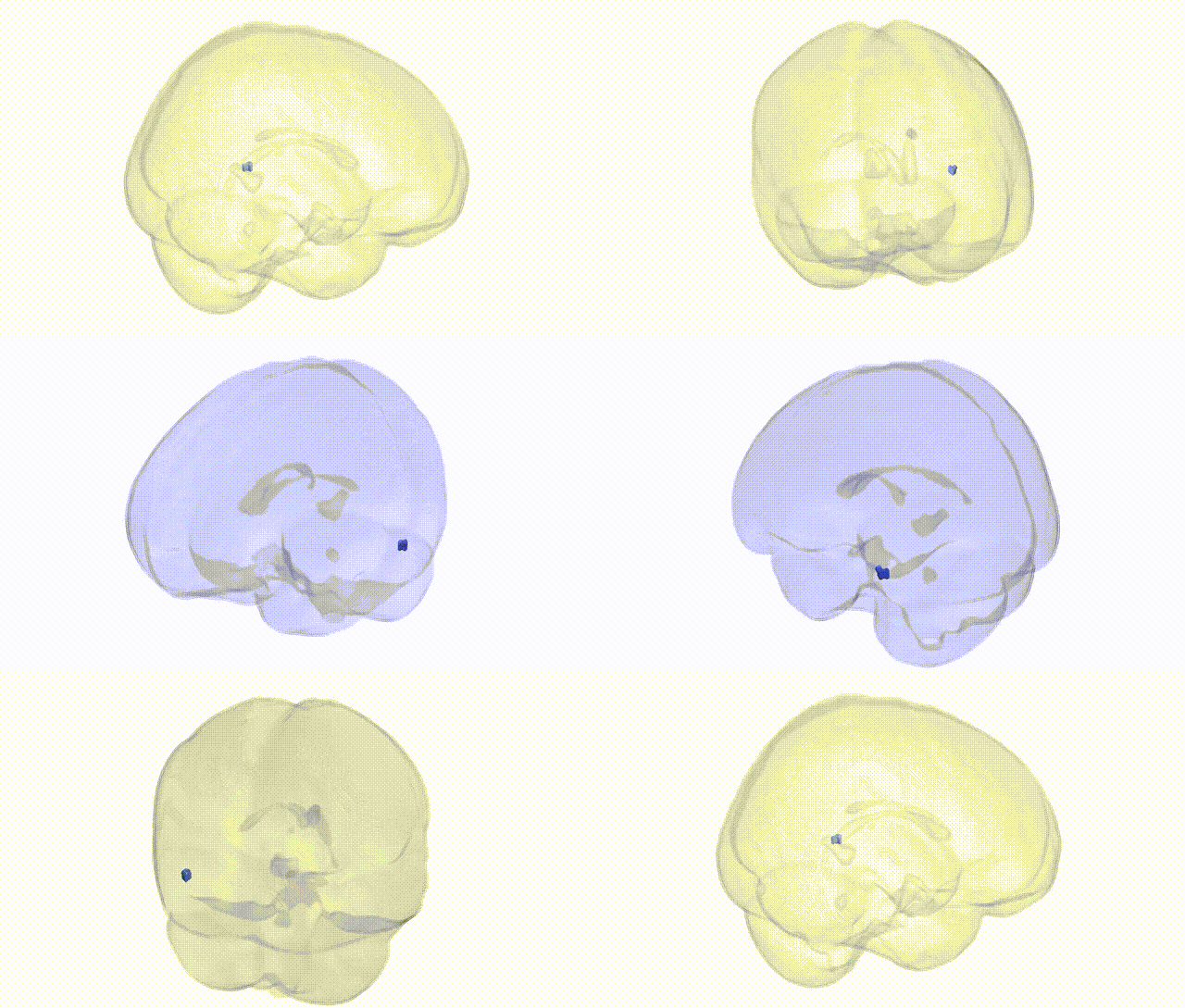
Full Quality Version: Download MP4
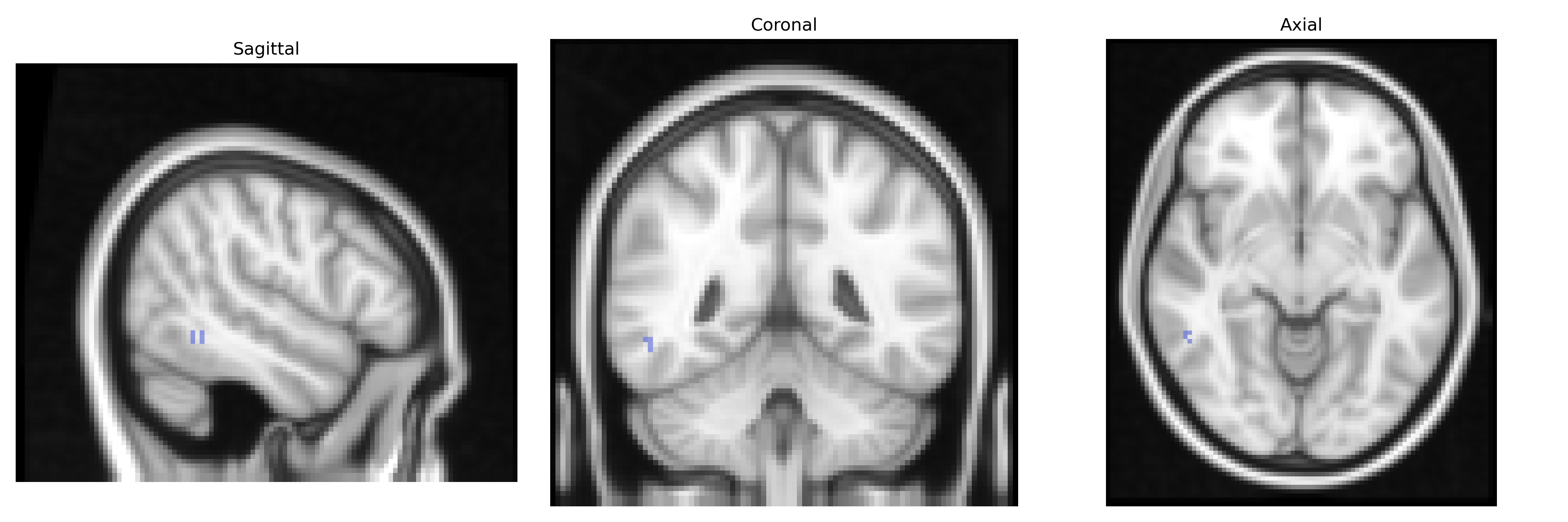
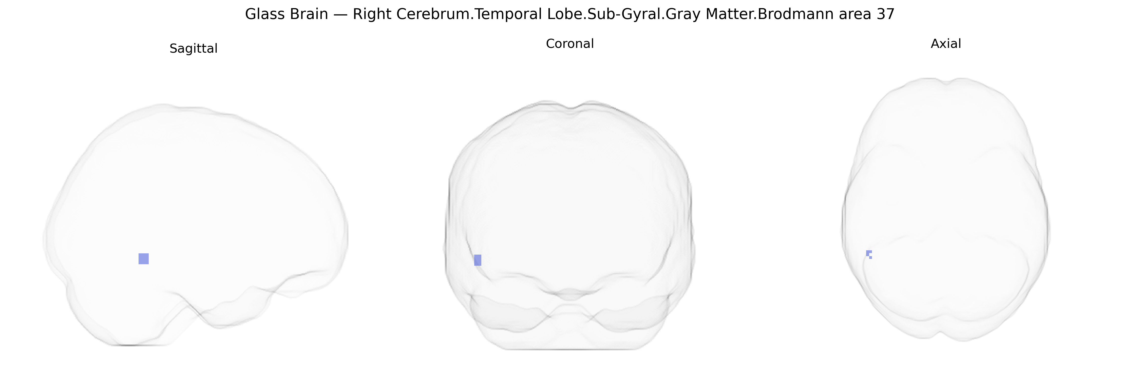
Wali Sidiqyar*, Gaurav Rudravaram*, Elyssa M. McMaster, Trent M. Schwartz, Adam M. Saunders, Kurt G. Schilling, Bennett A. Landman "Introducing SPINS: A Shared Public Visualization Library of Neuroanatomical Structures." Medical Imaging with Deep Learning- short paper
This resource is licensed under CC0 1.0 Universal (Public Domain).