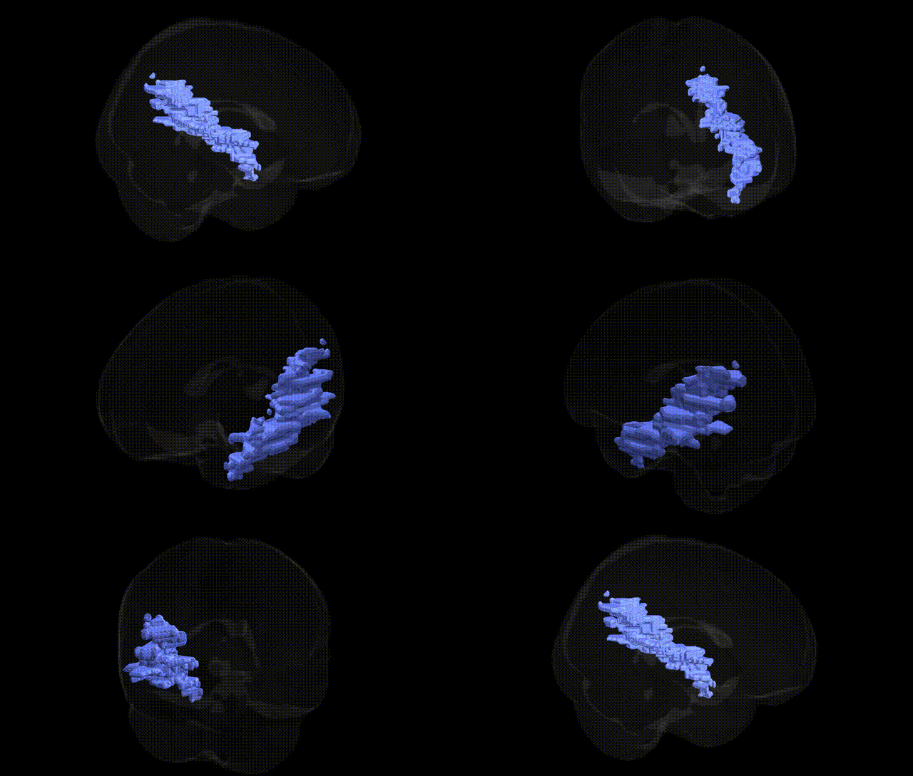
The bilateral Right Cerebrum.Temporal Lobe.Sub-Gyral.White Matter refers to deep white matter located beneath the cortical gyri of the right temporal lobe, encompassing association, commissural, and projection fibers that interconnect temporal cortical regions with other parts of the cerebrum and subcortical structures. These fiber tracts include portions of major pathways such as the inferior longitudinal fasciculus, arcuate fasciculus, and components of the temporal segment of the optic radiations, supporting functions related to auditory processing, language, memory, and higher-order visual integration. As a white matter compartment rather than a distinct nucleus or gyrus, this region serves primarily as a conduit for information flow between temporal cortical areas (e.g., superior, middle, and inferior temporal gyri) and distributed neural networks involved in perception, cognition, and emotion. There is no direct link for this specific sub-gyral white matter region; a related structure is the Temporal lobe.
Genetic associations involving the bilateral right temporal sub-gyral white matter (as defined in the Talairach 2 mm atlas) arise largely from imaging genetics and GWAS of regional white matter microstructure rather than from studies targeting this specific label. Variants in genes influencing myelination and axonal integrity (such as CNTNAP2, NRG1, MAG, MBP, and FKBP5) have been repeatedly linked to temporal lobe white matter properties in diffusion MRI studies, often in the context of schizophrenia, bipolar disorder, major depressive disorder, autism spectrum disorder, and anxiety-related traits. Large-scale GWAS of diffusion tensor imaging measures (e.g., mean diffusivity, fractional anisotropy) in temporal white matter tracts have identified multiple loci in neurodevelopmental and synaptic genes, including BDNF, cell-adhesion and axon-guidance genes, and immune-related loci (e.g., in the MHC region), with downstream associations to cognitive performance, language abilities, psychosis risk, and neuroticism. Temporal lobe white matter has been implicated in genetic risk for temporal lobe epilepsy, with variants in ion channel and synaptic genes (such as SCN1A, GABRA2, and LGI1) associated with structural and connectivity alterations that encompass sub-gyral regions. Additionally, APOE ε4 and other Alzheimer’s disease risk loci show effects on temporal white matter integrity, linking this region to genetic susceptibility for dementia and age-related cognitive decline, although the fine-grained Talairach sub-gyral label itself has not been the direct focus of most GWAS and is typically subsumed within broader temporal lobe or tract-based analyses.
Overview generated by GPT-4o (2026).
Region ID: 52
Hemisphere: bilateral
Atlas: Talairach labels 2mm
Full Quality Version: Download MP4
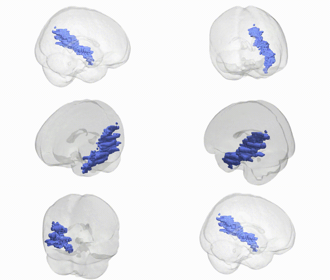
Full Quality Version: Download MP4
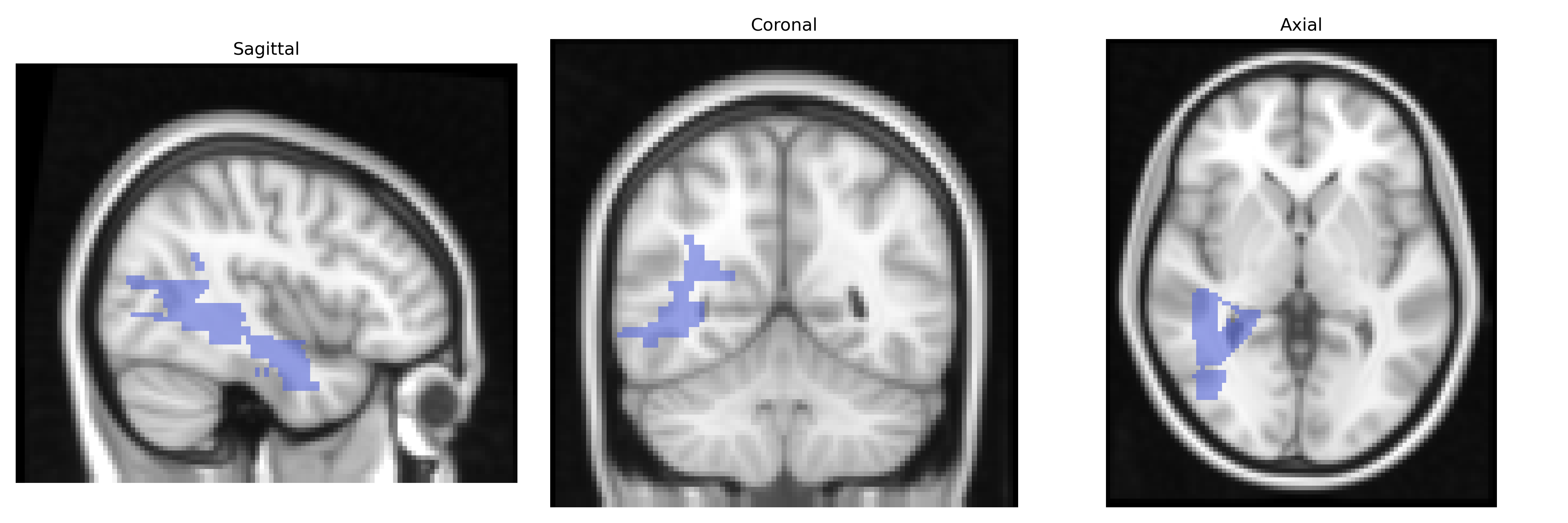
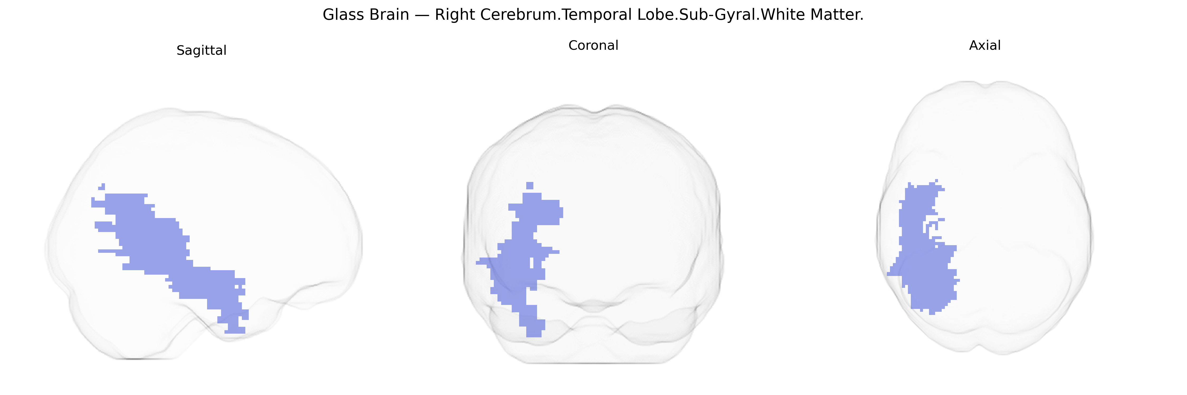
Wali Sidiqyar*, Gaurav Rudravaram*, Elyssa M. McMaster, Trent M. Schwartz, Adam M. Saunders, Kurt G. Schilling, Bennett A. Landman "Introducing SPINS: A Shared Public Visualization Library of Neuroanatomical Structures." Medical Imaging with Deep Learning- short paper
This resource is licensed under CC0 1.0 Universal (Public Domain).