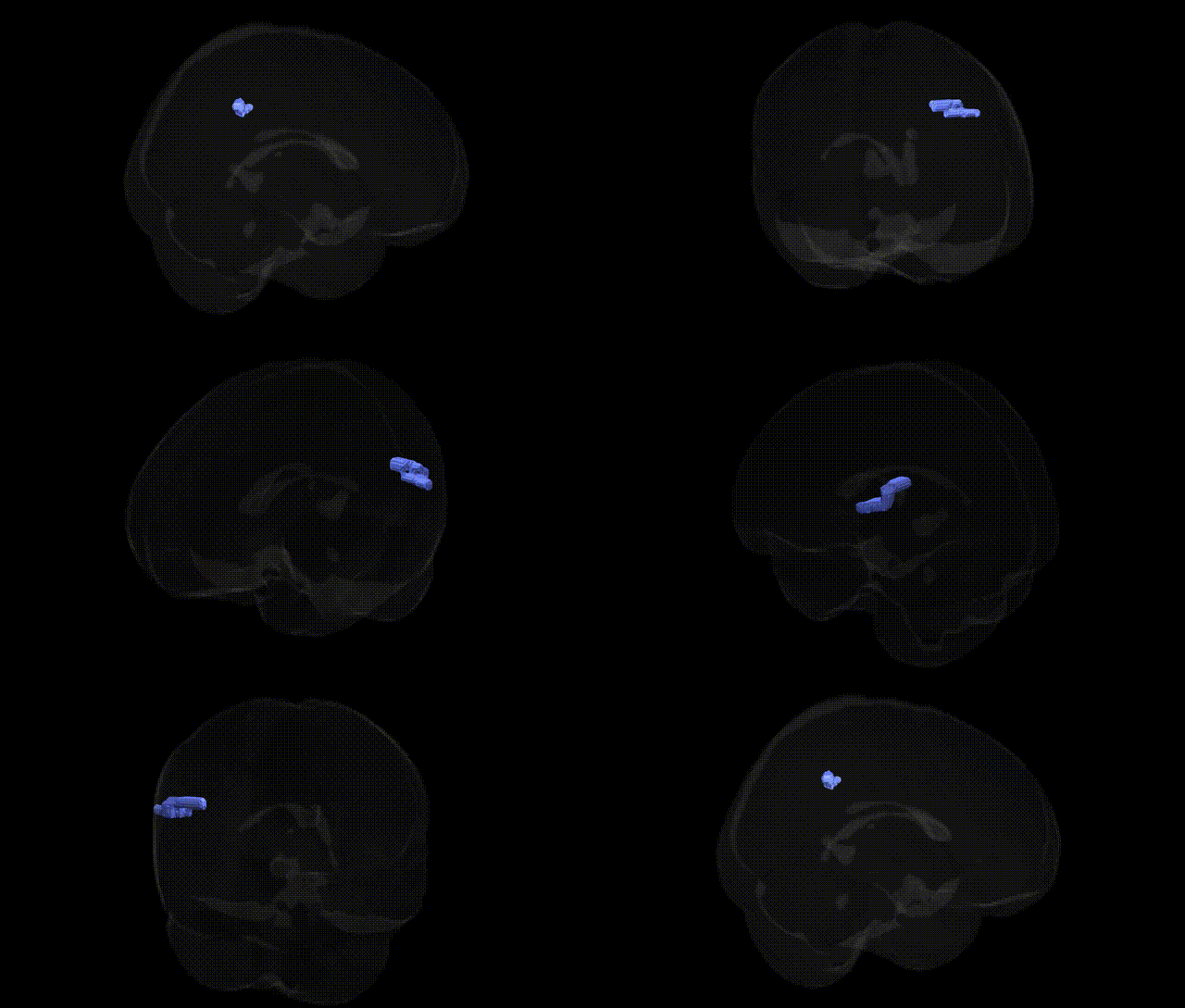
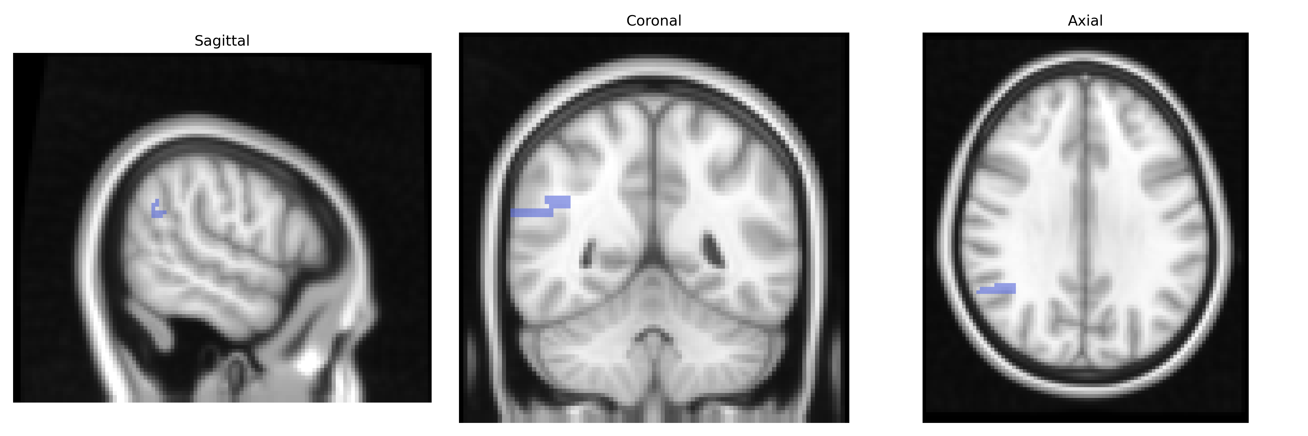
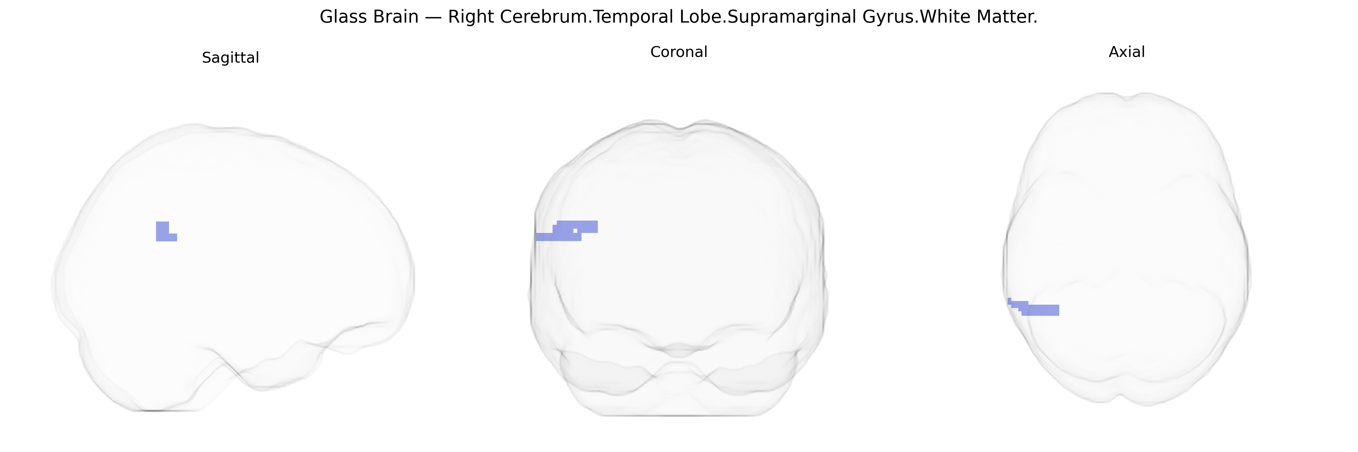
The bilateral Right Cerebrum.Temporal Lobe.Supramarginal Gyrus.White Matter corresponds to subcortical association fibers underlying the supramarginal gyrus, a key subdivision of the inferior parietal lobule located at the parietal–temporal junction. Although labeled under the temporal lobe in the Talairach 2 mm atlas, this region’s white matter consists largely of short and long-range corticocortical pathways interconnecting supramarginal cortex with adjacent parietal, temporal, and frontal areas, supporting multimodal integration of somatosensory, auditory, and language-related information. Functionally, these fibers contribute to phonological processing, articulation, sensorimotor integration for speech, reading, and aspects of spatial attention and body representation. There is no direct link for this exact white matter region; a related cortical structure is the Supramarginal gyrus.
Genetic associations for the bilateral Right Cerebrum.Temporal Lobe.Supramarginal Gyrus.White Matter region, as defined in the Talairach labels 2 mm atlas, are typically inferred from broader studies of the supramarginal gyrus, temporoparietal and perisylvian white matter, and diffusion MRI measures (e.g., fractional anisotropy) rather than from this exact parcel. GWAS of white matter microstructure have implicated loci near genes such as CACNA1C, ZNF804A, and NRG1, which are involved in synaptic function and myelination and have been associated with schizophrenia and bipolar disorder, showing effects in temporoparietal association fibers that traverse or border the supramarginal region. Variants in APOE (particularly ε4) and CLU linked to Alzheimer’s disease have been associated with reduced integrity of temporoparietal white matter, including tracts connecting the supramarginal gyrus with temporal and frontal language and attentional networks. GWAS of reading and language-related traits, including dyslexia, have identified risk variants near DCDC2, KIAA0319, and ROBO1, whose effects on perisylvian white matter pathways (e.g., arcuate/superior longitudinal fasciculus) overlap with supramarginal gyrus white matter and influence phonological processing and literacy. In autism spectrum conditions and social cognition traits, polygenic risk scores and candidate gene studies involving synaptic and neurodevelopmental genes (such as CNTNAP2, SHANK3, and NRXN1) have been linked to altered microstructure in temporoparietal junction and supramarginal-associated tracts, supporting a genetic contribution to social and language integration functions subserved by this region. Overall, genetic findings converge on neurodevelopmental, synaptic, and myelin-related pathways that influence the structure and connectivity of supramarginal gyrus white matter and its involvement in language, reading, social cognition, and susceptibility to major psychiatric and neurodegenerative disorders.
Overview generated by GPT-4o (2026).
Region ID: 871
Hemisphere: bilateral
Atlas: Talairach labels 2mm
Full Quality Version: Download MP4
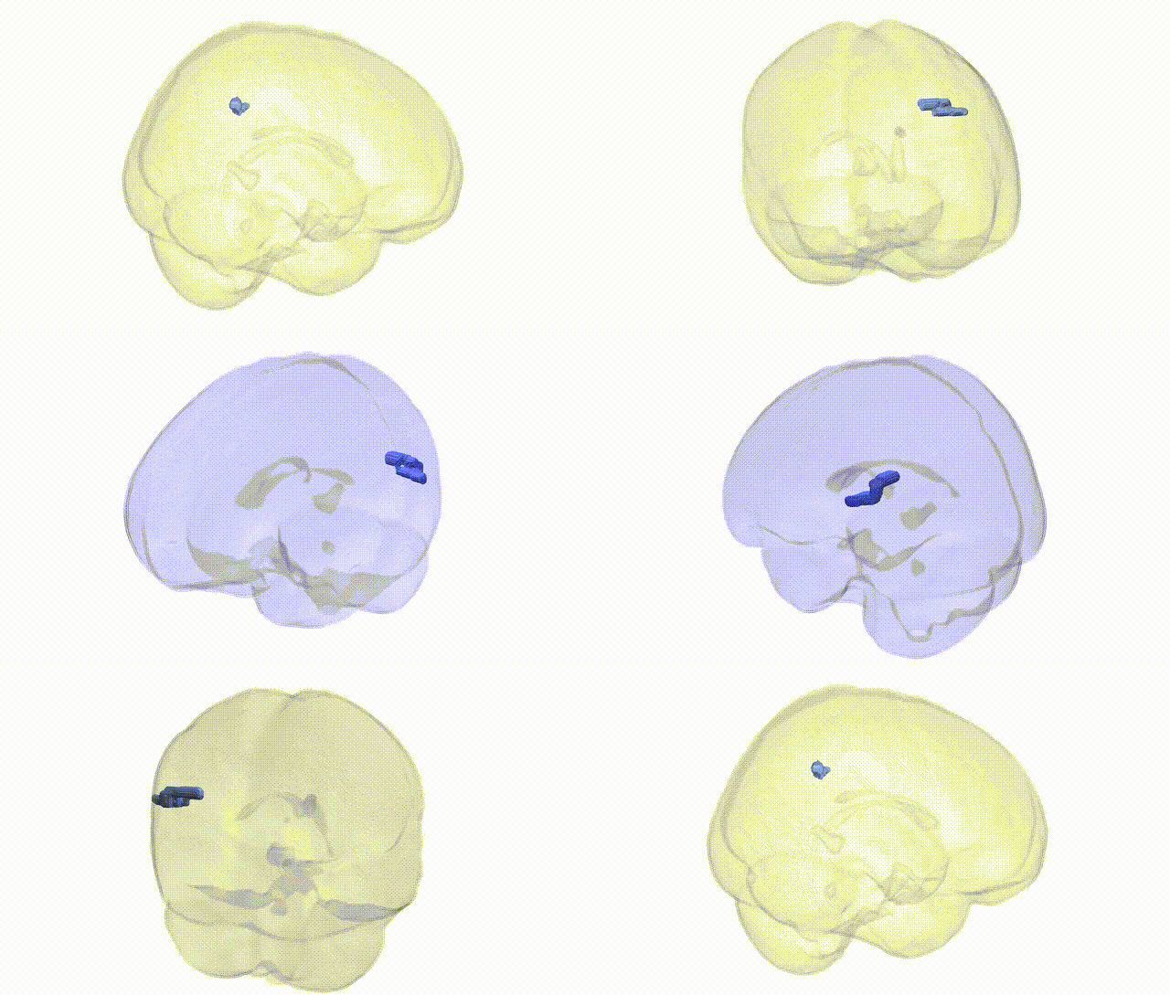
Full Quality Version: Download MP4
Wali Sidiqyar*, Gaurav Rudravaram*, Elyssa M. McMaster, Trent M. Schwartz, Adam M. Saunders, Kurt G. Schilling, Bennett A. Landman "Introducing SPINS: A Shared Public Visualization Library of Neuroanatomical Structures." Medical Imaging with Deep Learning- short paper
This resource is licensed under CC0 1.0 Universal (Public Domain).