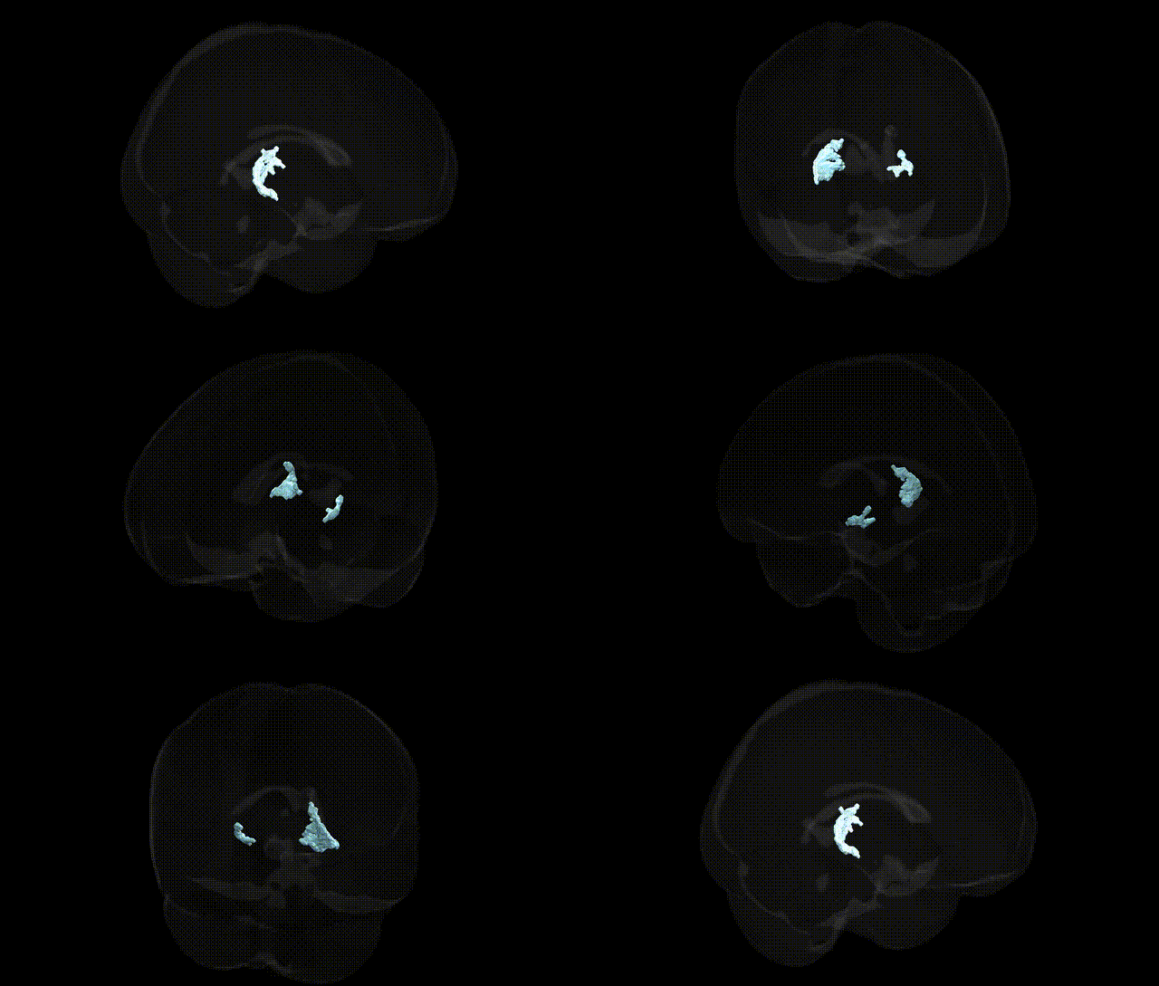
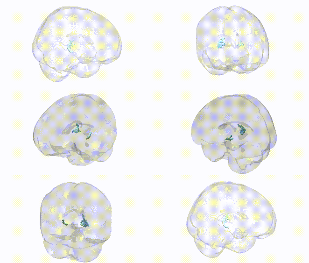
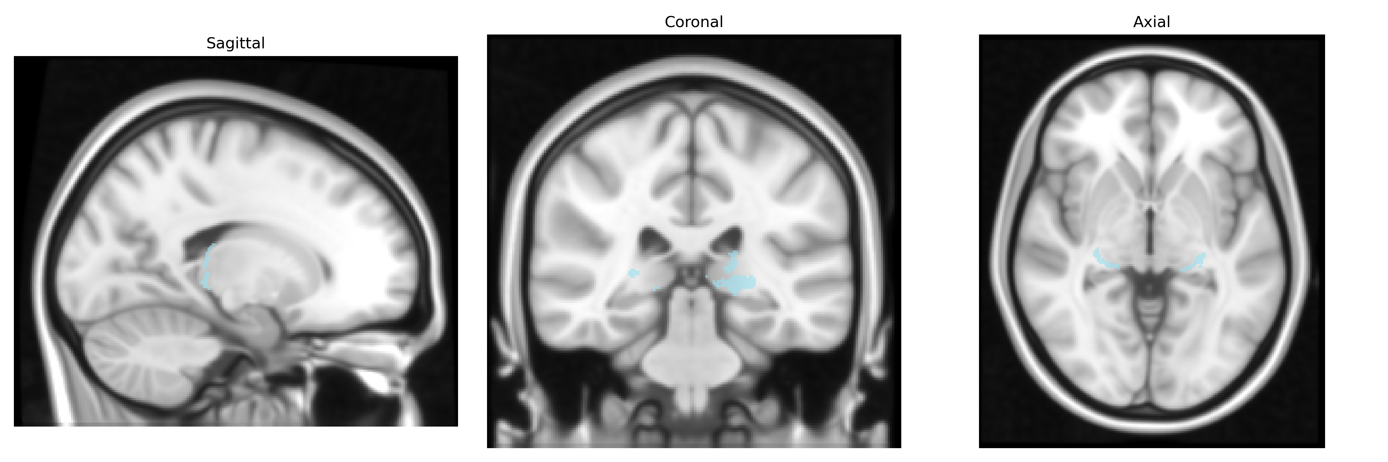
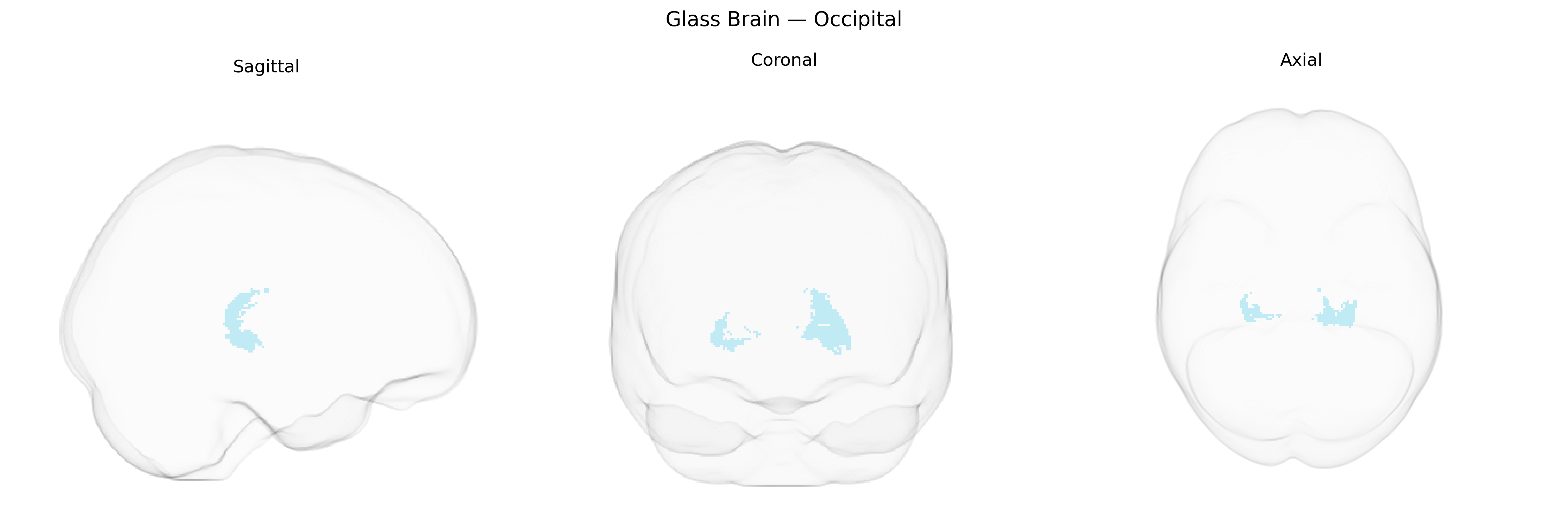
The bilateral occipital thalamic region in the Thalamus maxprob thr25 1mm atlas corresponds primarily to lateral geniculate and adjacent pulvinar nuclei that relay and modulate visual information from the retina to the primary and associative visual cortices. These nuclei receive topographically organized retinal input via the optic tract and project predominantly to the occipital lobe, particularly the calcarine (striate) cortex and surrounding extrastriate areas, thereby supporting conscious visual perception, spatial processing, and aspects of visuomotor integration. Functionally, this region participates in parallel visual pathways (e.g., magnocellular and parvocellular channels), contributes to contrast sensitivity, motion and color processing, and interacts with cortical feedback loops that refine visual attention and representation. There is no direct link for this exact atlas-defined region; a closely related structure is the Lateral geniculate nucleus.
Genetic associations involving bilateral occipital regions overlapping or functionally connected with thalamic nuclei (as defined in the Thalamus maxprob thr25 1 mm atlas) primarily emerge from large-scale imaging–genetics and GWAS studies linking thalamocortical and visual network structure to polygenic variation. Several loci influencing cortical surface area and thickness in occipital cortex—such as variants near genes involved in neurodevelopment and synaptic function (e.g., MAPT, GRIN2B, and other glutamatergic and axon guidance genes)—have shown associations with occipital morphology or visual processing measures, and some of these loci also affect thalamic volumes or connectivity, suggesting coordinated genetic control of thalamus–occipital circuits. GWAS of disorders with pronounced visual or occipital involvement, including migraine with aura, occipital lobe epilepsy, and certain subtypes of schizophrenia and major depression, have identified risk variants in genes affecting synaptic transmission, myelination, and neurovascular regulation, which in turn correlate with altered occipital activation and thalamocortical signaling. In neurodegenerative and neurodevelopmental conditions, such as Alzheimer’s disease, multiple sclerosis, and autism spectrum disorder, polygenic risk scores have been associated with both occipital structural changes and thalamic network alterations, implicating shared genetic influences on sensory integration, visual perception, and higher-order thalamocortical processing in the bilateral occipital–thalamic system.
Overview generated by GPT-4o (2026).
Region ID: 3
Hemisphere: bilateral
Atlas: Thalamus maxprob thr25 1mm
Full Quality Version: Download MP4
Full Quality Version: Download MP4
Wali Sidiqyar*, Gaurav Rudravaram*, Elyssa M. McMaster, Trent M. Schwartz, Adam M. Saunders, Kurt G. Schilling, Bennett A. Landman "Introducing SPINS: A Shared Public Visualization Library of Neuroanatomical Structures." Medical Imaging with Deep Learning- short paper
This resource is licensed under CC0 1.0 Universal (Public Domain).