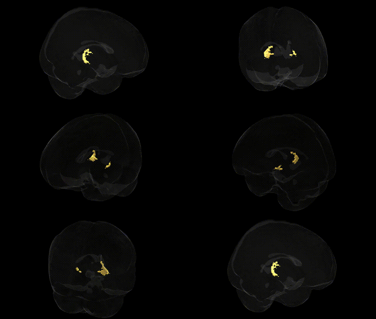
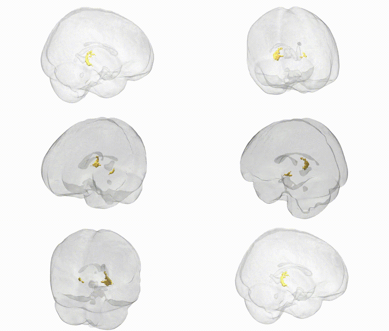
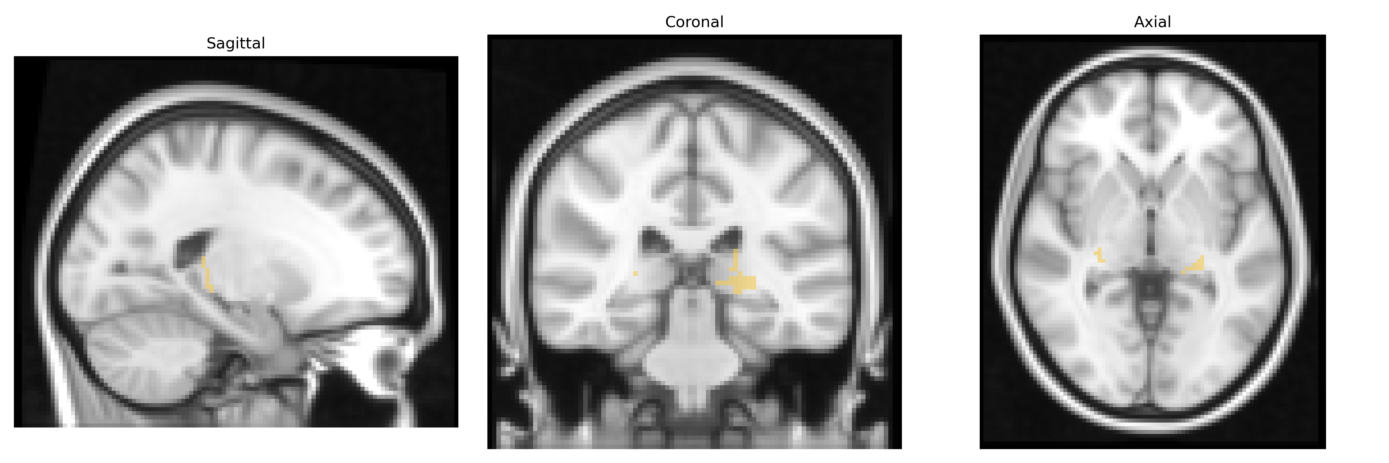
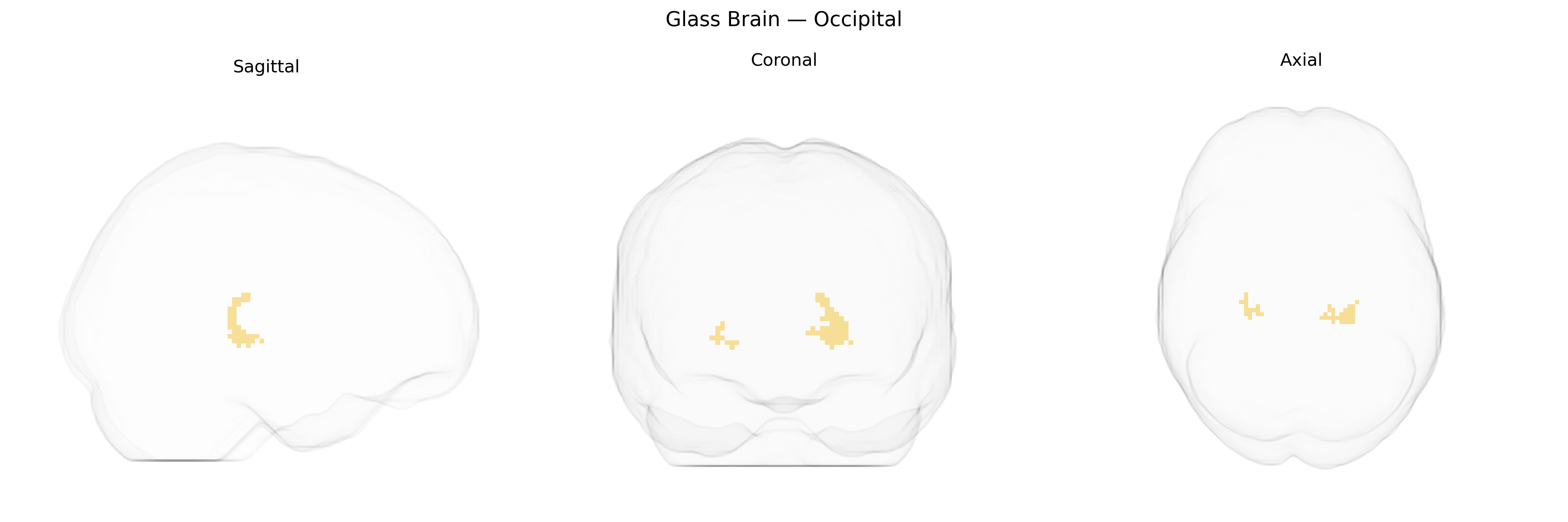
The bilateral Occipital region in the Thalamus maxprob thr25 2mm atlas corresponds primarily to thalamic nuclei projecting to and associated with the occipital cortex, particularly those involved in visual information processing. Functionally, these thalamic territories participate in relaying and modulating visual signals from subcortical structures (notably the lateral geniculate and adjacent visual relay nuclei) to the primary and secondary visual cortices, contributing to visual perception, spatial orientation, and integration of visual input with higher-order cortical areas. This occipital-related thalamic group is integral to the cortico-thalamo-cortical loops that underlie conscious visual experience and visual attention, and it interacts with both dorsal and ventral visual stream pathways. There is no direct Wikipedia article for this atlas-defined “Thalamic Occipital” region; a closely related structure is the occipital lobe: Occipital lobe.
Genetic associations involving the bilateral occipital region in the context of thalamocortical networks, including areas mapped in the Thalamus maxprob thr25 2 mm Atlas, point to a role of common variants in synaptic signaling, neurodevelopmental, and visual-processing pathways. GWAS of brain cortical and subcortical morphology (e.g., ENIGMA and UK Biobank studies) have identified loci near genes such as TCF4, EPHB4, PAX6, GRIN2A, and KIAA0586 that influence occipital cortical thickness, surface area, and thalamic volume, implicating glutamatergic neurotransmission, axon guidance, and neurogenesis in structural variability of occipital–thalamic circuits. Polygenic risk for schizophrenia, bipolar disorder, and major depressive disorder is associated with altered occipital and thalamic structure and connectivity, while autism spectrum disorder and ADHD GWAS highlight genes involved in sensory integration and attention that modulate activity in occipital–thalamic pathways. Variants in visual system–related genes (e.g., associated with refractive error, visual acuity, and migraine with aura) have also been linked to occipital cortical measures, and migraine GWAS repeatedly implicate thalamic and occipital functional networks, suggesting a shared genetic basis for visual aura and sensory processing abnormalities. Collectively, these findings support the bilateral occipital region within thalamocortical circuits as a genetically influenced hub for visual perception, higher-order cognition, and vulnerability to neuropsychiatric and neurological disorders.
Overview generated by GPT-4o (2026).
Region ID: 3
Hemisphere: bilateral
Atlas: Thalamus maxprob thr25 2mm
Full Quality Version: Download MP4
Full Quality Version: Download MP4
Wali Sidiqyar*, Gaurav Rudravaram*, Elyssa M. McMaster, Trent M. Schwartz, Adam M. Saunders, Kurt G. Schilling, Bennett A. Landman "Introducing SPINS: A Shared Public Visualization Library of Neuroanatomical Structures." Medical Imaging with Deep Learning- short paper
This resource is licensed under CC0 1.0 Universal (Public Domain).