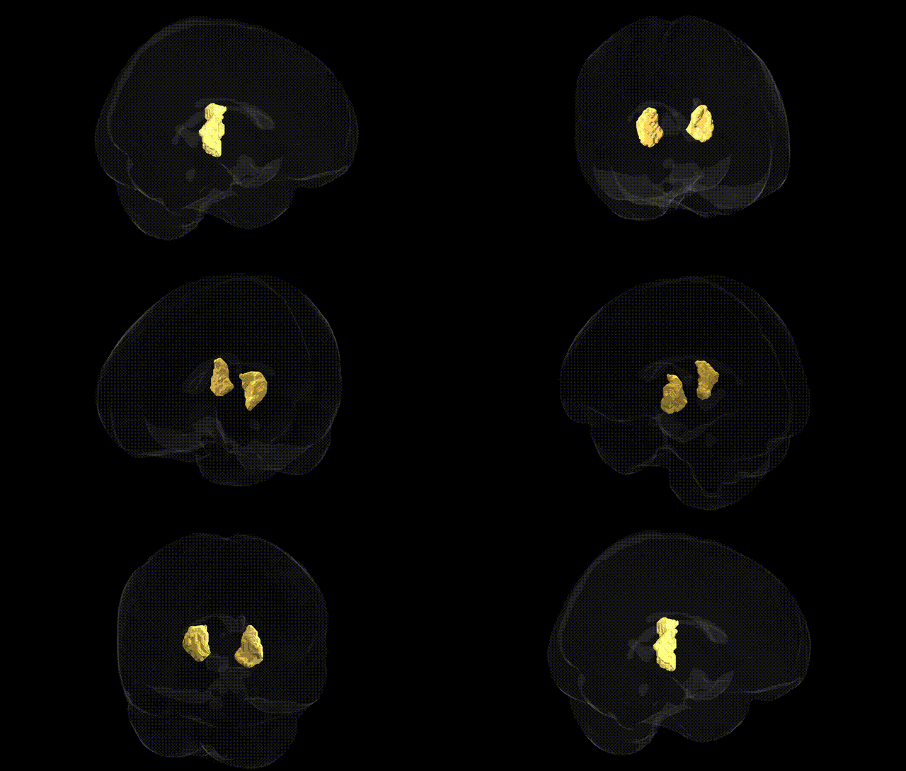
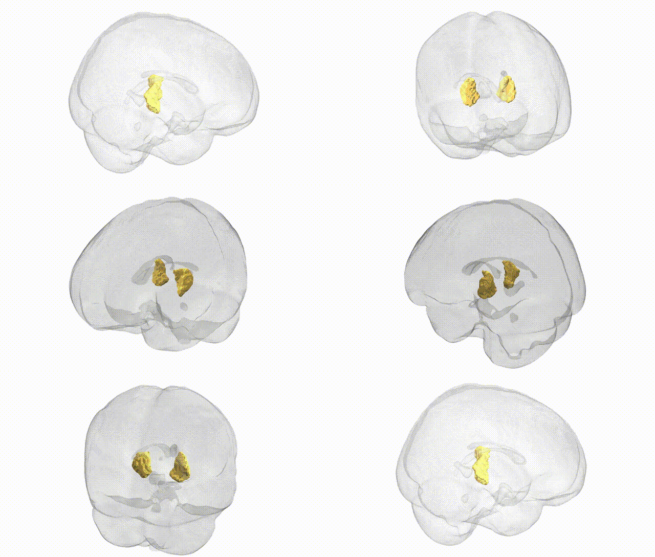
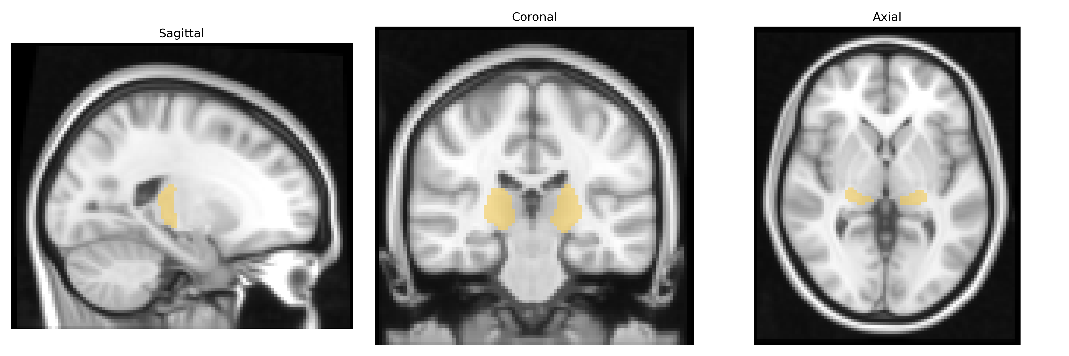
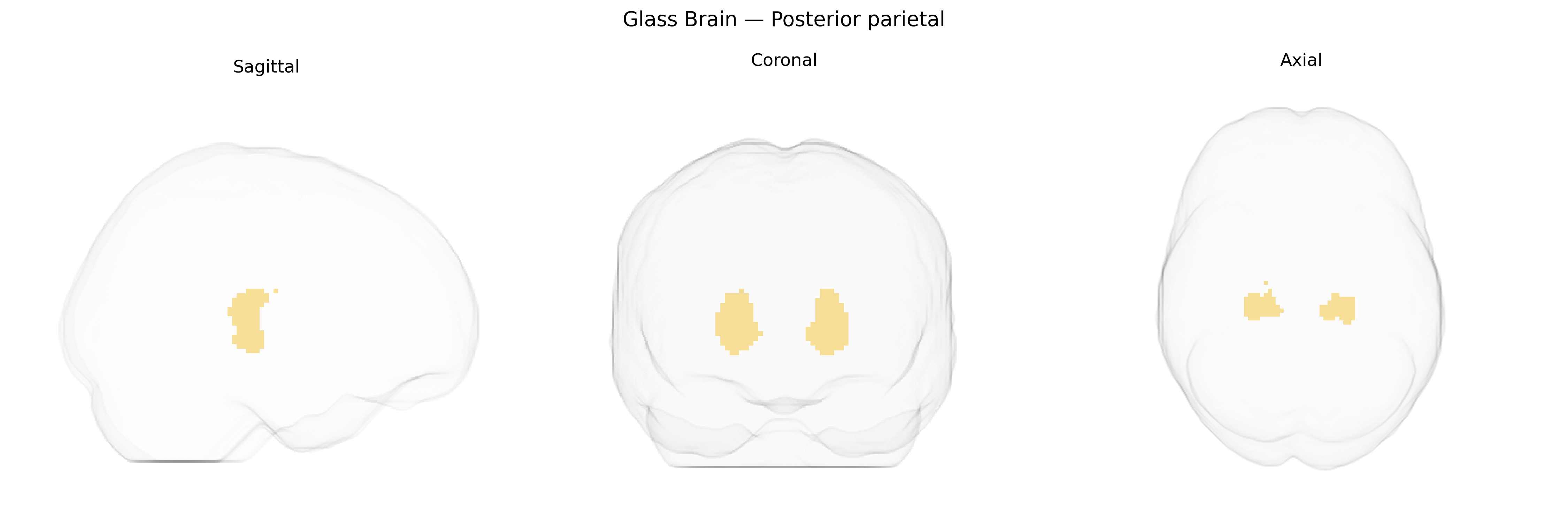
The bilateral posterior parietal region in the Thalamus maxprob thr25 2 mm atlas corresponds to thalamic nuclei that predominantly project to and modulate activity in the posterior parietal cortex, a multimodal association area involved in integrating somatosensory, visual, and proprioceptive inputs to support spatial perception, attention, sensorimotor transformations, and goal-directed behavior. These thalamic territories, which include higher-order relay and association nuclei, contribute to the coordination of visuospatial processing, body schema representation, and the planning and guidance of limb and eye movements, particularly in tasks requiring spatial attention and the transformation of sensory coordinates into motor commands. Dysfunction in this circuit is associated with deficits such as hemispatial neglect, impaired visuomotor coordination, and disruptions of complex spatial cognition. No direct Wikipedia article exists for the “posterior parietal thalamic region” as defined by this atlas; a closely related cortical target region is the Posterior parietal cortex.
Genetic associations involving the bilateral posterior parietal region, as defined via thalamocortical connectivity in the Thalamus maxprob thr25 2mm atlas, largely emerge from imaging genetics and GWAS that link parietal structure and function to cognitive, neurodevelopmental, and psychiatric traits. Variants in genes implicated in synaptic plasticity and neurodevelopment, such as BDNF (e.g., Val66Met), COMT, and KIBRA (WWC1), have been associated with parietal activation and connectivity during working memory, attention, and episodic memory tasks, while large-scale GWAS of cortical thickness and surface area (e.g., ENIGMA and UK Biobank studies) have identified loci in pathways related to neuronal differentiation and axon guidance that influence parietal morphology. Polygenic risk for schizophrenia, autism spectrum disorder, and attention-deficit/hyperactivity disorder shows associations with altered parietal structure and thalamocortical connectivity, and Alzheimer’s disease risk genes such as APOE have been linked to posterior parietal atrophy and hypometabolism, particularly in the context of default-mode and attentional network dysfunction. Additional GWAS of general cognitive ability, educational attainment, and mathematical/visuospatial skills have reported that genetic variants contributing to these traits are associated with functional and structural metrics in the posterior parietal cortex, consistent with its role in higher-order cognition and sensory integration.
Overview generated by GPT-4o (2026).
Region ID: 6
Hemisphere: bilateral
Atlas: Thalamus maxprob thr25 2mm
Full Quality Version: Download MP4
Full Quality Version: Download MP4
Wali Sidiqyar*, Gaurav Rudravaram*, Elyssa M. McMaster, Trent M. Schwartz, Adam M. Saunders, Kurt G. Schilling, Bennett A. Landman "Introducing SPINS: A Shared Public Visualization Library of Neuroanatomical Structures." Medical Imaging with Deep Learning- short paper
This resource is licensed under CC0 1.0 Universal (Public Domain).