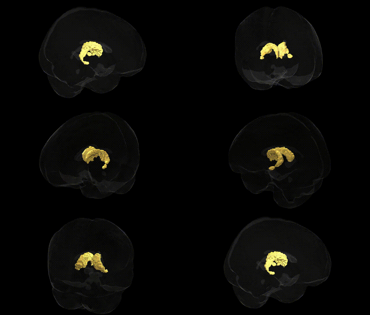
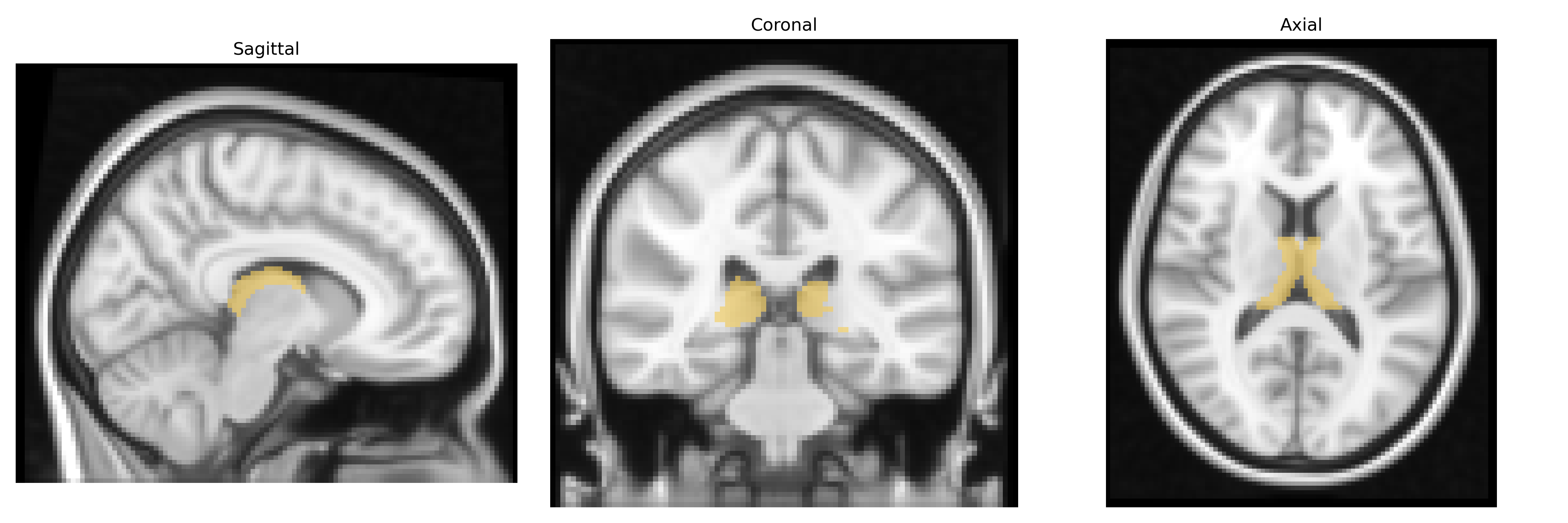
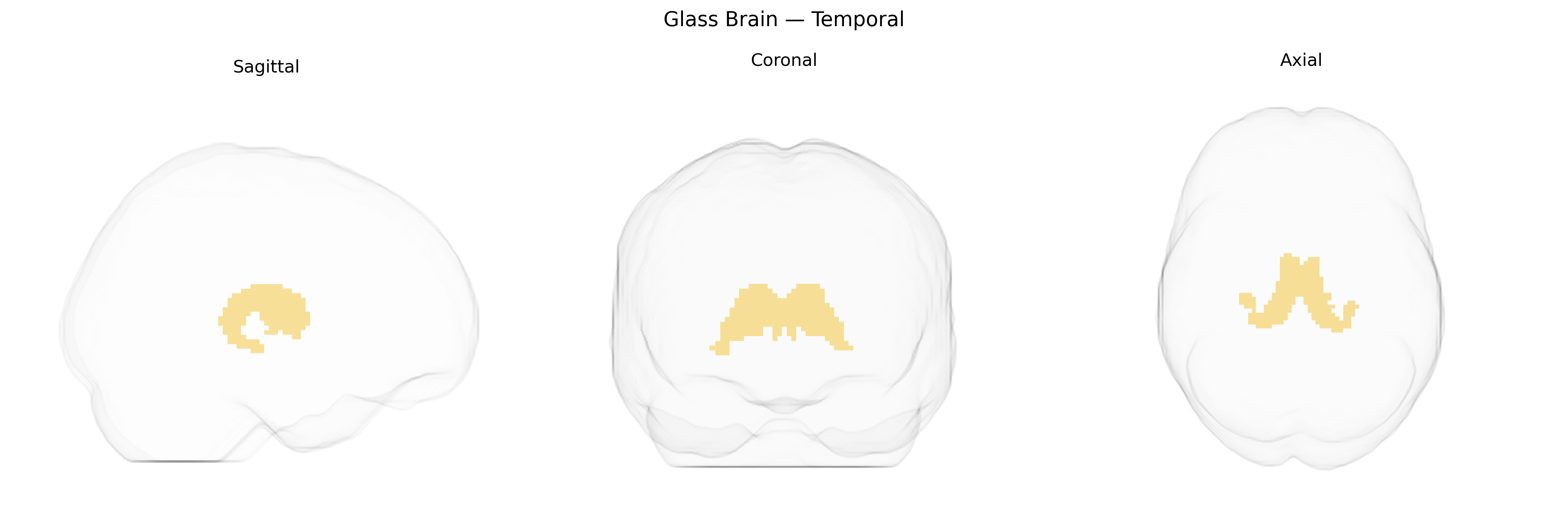
The bilateral temporal region in the Thalamus maxprob thr25 2mm atlas principally corresponds to thalamic nuclei projecting to the temporal cortex, including components of the medial geniculate nucleus and adjacent nuclei involved in auditory processing, multimodal integration, and aspects of memory and emotion. These thalamic territories relay auditory information from the midbrain (inferior colliculus) to primary and associative auditory cortices, participate in higher-order auditory perception such as sound localization and complex pattern recognition, and contribute to limbic circuits that influence emotional valence and mnemonic encoding of auditory stimuli. Functionally, this region acts as a key hub linking sensory input to temporal lobe structures like the hippocampus and amygdala, supporting integration of auditory signals with contextual, emotional, and episodic memory processes. There is no direct Wikipedia article for this exact atlas-defined “bilateral temporal” thalamic region; a closely related structure is the medial geniculate nucleus: Medial geniculate nucleus.
The bilateral temporal region of the thalamus, as delineated in the Thalamus maxprob thr25 2mm Atlas, has been implicated in several genetic and GWAS-based associations, largely through studies of thalamic and temporo-thalamic connectivity rather than this subregion in isolation. Variants in genes involved in neurodevelopment and synaptic function—such as CACNA1C, GRIN2A, and CNTNAP2—have been linked to alterations in thalamic volume and functional coupling with temporal cortices, with downstream associations to schizophrenia, bipolar disorder, and major depressive disorder. Polygenic risk scores for schizophrenia and autism spectrum disorder have shown relationships with thalamocortical circuit structure and function, including pathways connecting the thalamus to temporal and auditory association areas, aligning with clinical findings of auditory hallucinations and language-processing deficits. GWAS of brain imaging phenotypes (e.g., ENIGMA, UK Biobank) report that common variants near genes involved in neuronal migration, axon guidance, and myelination (such as BDNF, NRG1, and multiple loci in the major histocompatibility complex region) are associated with variability in thalamic morphology and temporal-lobe–related networks, which in turn correlate with cognitive traits, including verbal memory, language ability, and general intelligence. Additionally, genetic risk for epilepsy, especially temporal lobe epilepsy, has been connected to disrupted thalamotemporal circuitry, with loci in GABAA receptor genes and ion channel genes contributing to seizure susceptibility through altered excitatory–inhibitory balance in these pathways.
Overview generated by GPT-4o (2026).
Region ID: 7
Hemisphere: bilateral
Atlas: Thalamus maxprob thr25 2mm
Full Quality Version: Download MP4
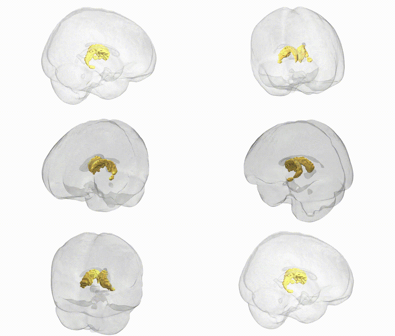
Full Quality Version: Download MP4
Wali Sidiqyar*, Gaurav Rudravaram*, Elyssa M. McMaster, Trent M. Schwartz, Adam M. Saunders, Kurt G. Schilling, Bennett A. Landman "Introducing SPINS: A Shared Public Visualization Library of Neuroanatomical Structures." Medical Imaging with Deep Learning- short paper
This resource is licensed under CC0 1.0 Universal (Public Domain).