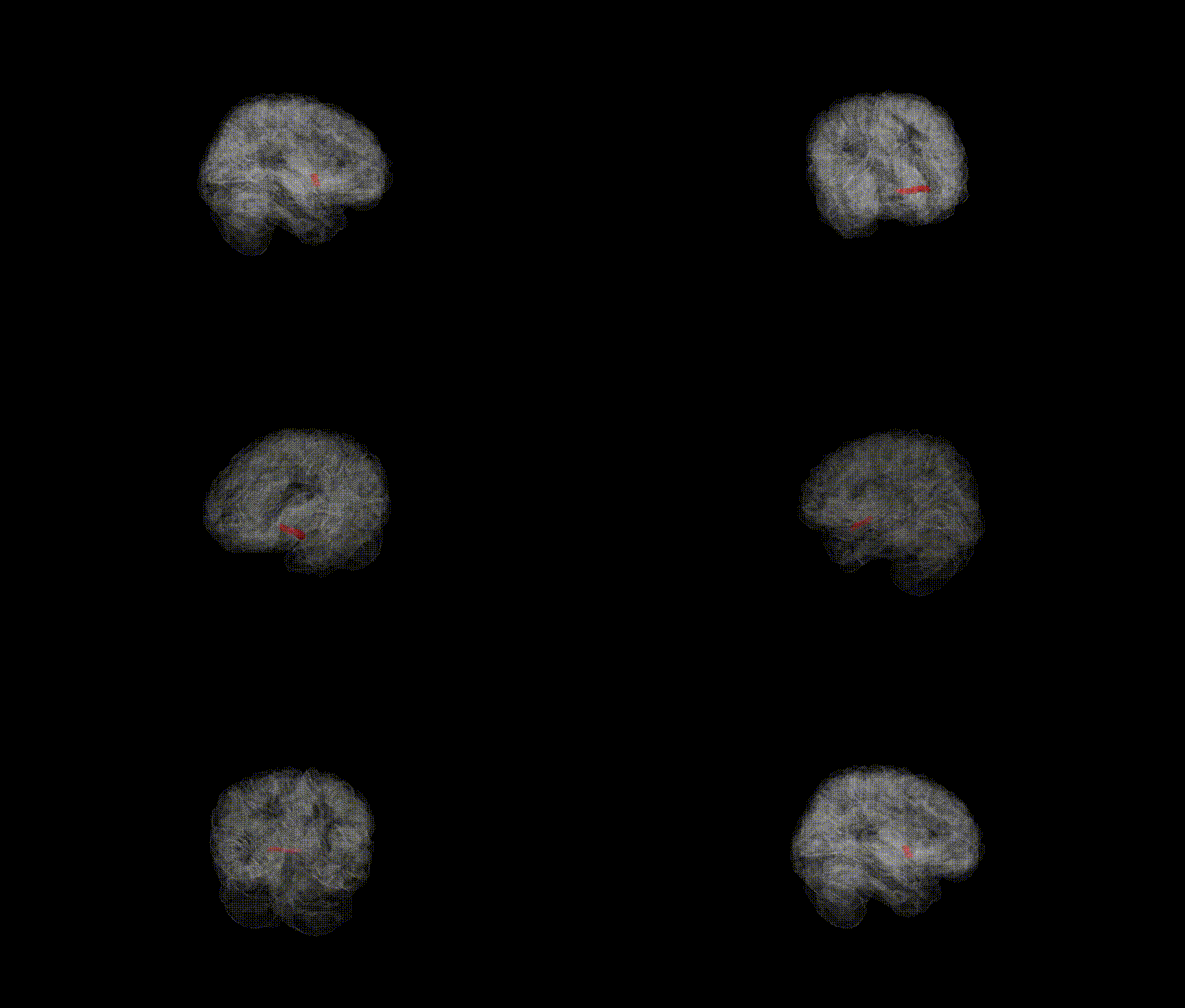
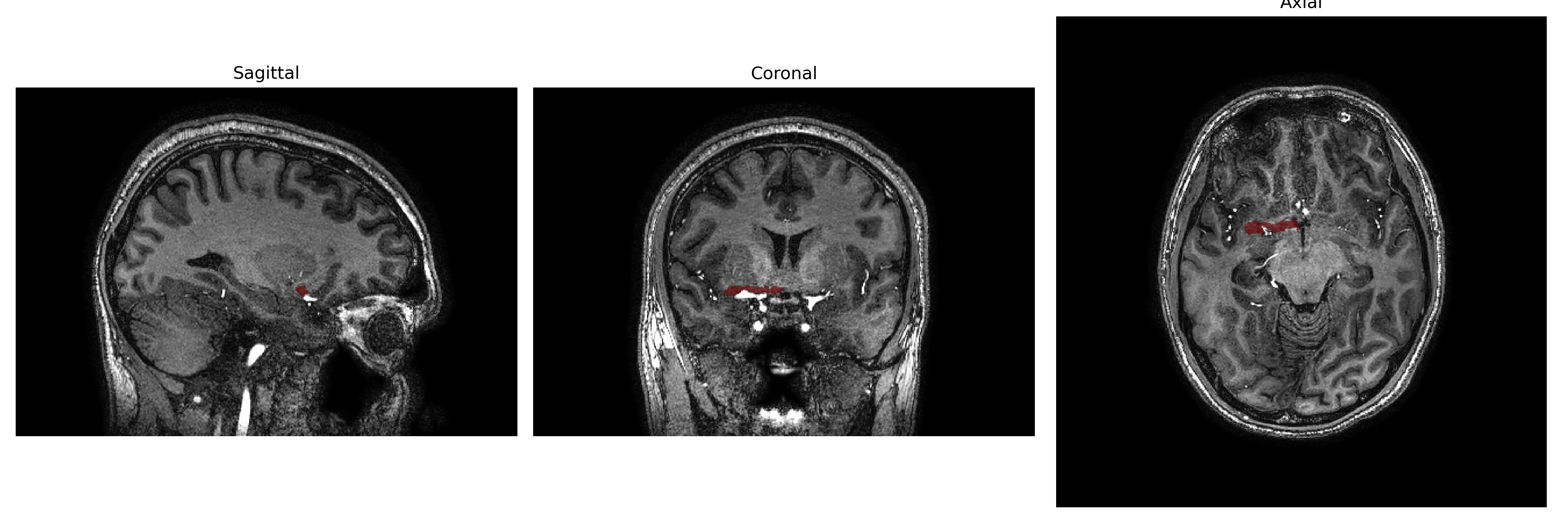
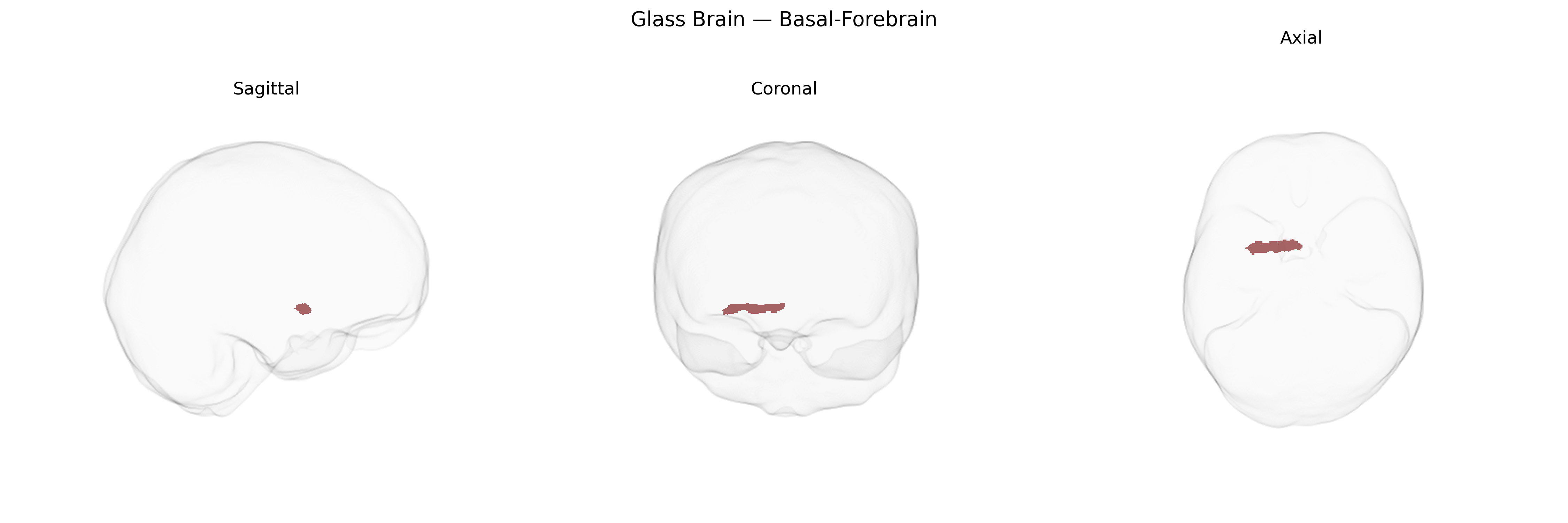
The Right Basal Forebrain in the brainCOLOR Atlas refers to the right-lateralized component of the basal forebrain, a collection of subcortical structures located in the ventral telencephalon anterior to the hypothalamus and below the anterior commissure. It includes major cholinergic nuclei such as the nucleus basalis of Meynert, medial septal nucleus, and diagonal band nuclei, which provide widespread neuromodulatory projections to the cerebral cortex, hippocampus, and amygdala. Functionally, this region is critically involved in regulating arousal, attention, learning, and memory through acetylcholine release, and it interacts with limbic and prefrontal networks to modulate cognitive flexibility and emotional processing. Degeneration of basal forebrain cholinergic neurons is a hallmark of neurodegenerative diseases, particularly Alzheimer’s disease and other dementias. There is no direct link for “Right Basal Forebrain”; a closely related structure is the Basal forebrain.
The Right Basal-Forebrain region, as defined in the brainCOLOR Atlas, encompasses cholinergic and limbic-associated nuclei whose structure and function show heritable variation and have been implicated in several genetic and GWAS findings, though often at the level of broader basal forebrain, medial septal, or nucleus basalis territories rather than this parcel alone. Imaging–genetics studies indicate moderate to high SNP-based heritability for basal forebrain volume and cholinergic system integrity, with common variants in genes related to cholinergic signaling (e.g., CHRNA7, CHRM2), neurotrophic factors (e.g., BDNF), and synaptic plasticity influencing measures of basal forebrain structure or activity. Large neuroimaging GWAS consortia (e.g., ENIGMA, UK Biobank-based analyses) have linked variants in and near APOE, CLU, BIN1, and PICALM—canonical Alzheimer’s disease risk genes—to reduced basal forebrain and adjacent medial temporal volumes, consistent with the region’s early degeneration in Alzheimer-type neurodegeneration. Genetic risk for schizophrenia, major depressive disorder, and anxiety-related traits has been associated with altered functional connectivity and microstructure in basal forebrain and limbic–striatal circuits, although these associations are typically reported at network or subcortical-composite levels rather than as right basal-forebrain-specific effects. Polygenic risk scores for cognitive performance and educational attainment show positive associations with basal forebrain volume and cholinergic system markers, while polygenic risk for Alzheimer’s disease tends to show the opposite pattern, underscoring a convergent role for this region as a genetically influenced hub in memory, attention, and neuropsychiatric vulnerability, even though direct GWAS hits explicitly localized to the “Right Basal-Forebrain” parcel of the brainCOLOR Atlas remain limited.
Overview generated by GPT-4o (2026).
Region ID: 23
Hemisphere: Right
Atlas: brainCOLOR
Full Quality Version: Download MP4
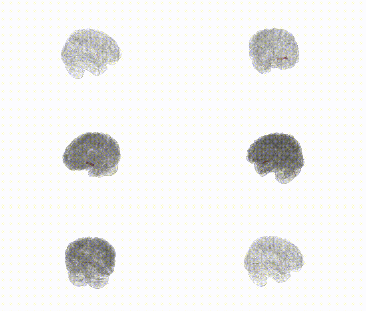
Full Quality Version: Download MP4
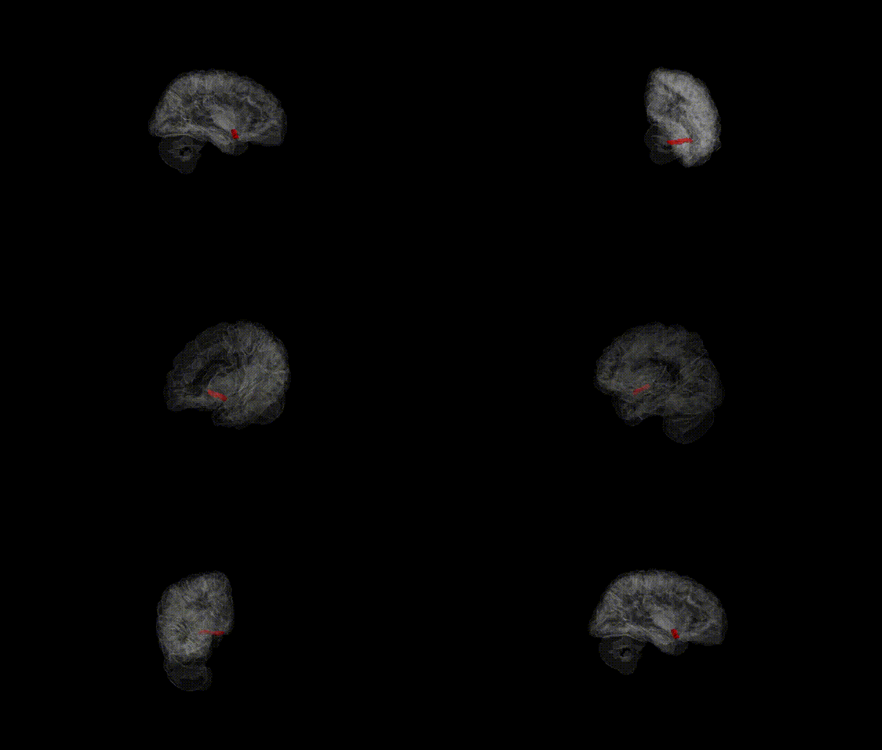
Full Quality Version: Download MP4
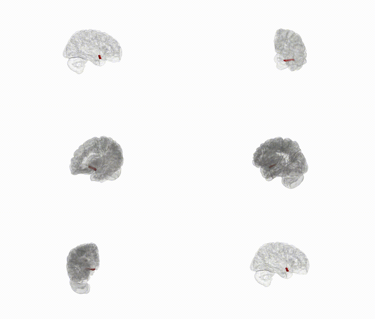
Full Quality Version: Download MP4
Wali Sidiqyar*, Gaurav Rudravaram*, Elyssa M. McMaster, Trent M. Schwartz, Adam M. Saunders, Kurt G. Schilling, Bennett A. Landman "Introducing SPINS: A Shared Public Visualization Library of Neuroanatomical Structures." Medical Imaging with Deep Learning- short paper
This resource is licensed under CC0 1.0 Universal (Public Domain).