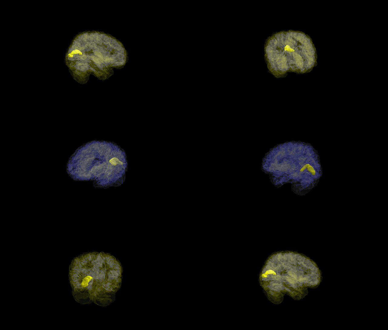
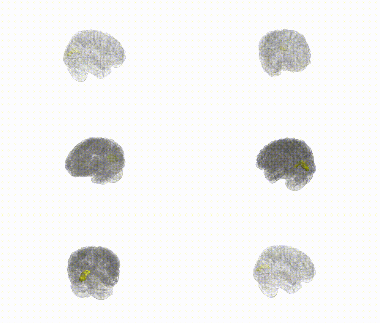
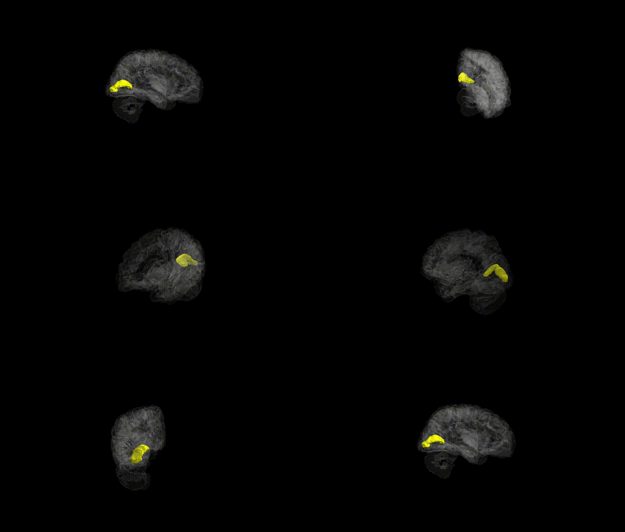
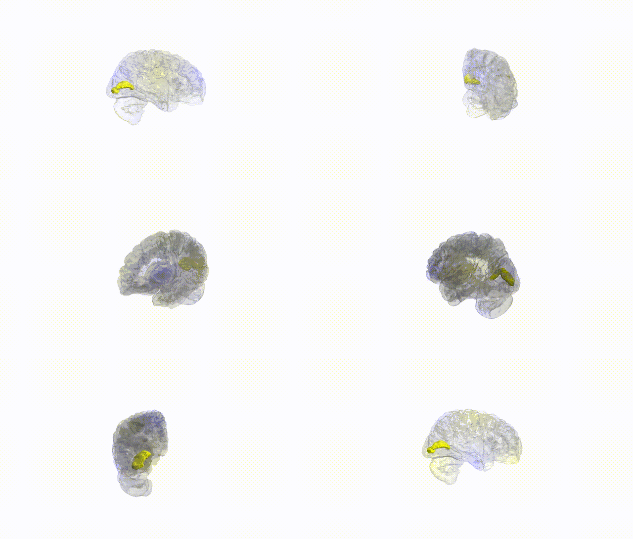
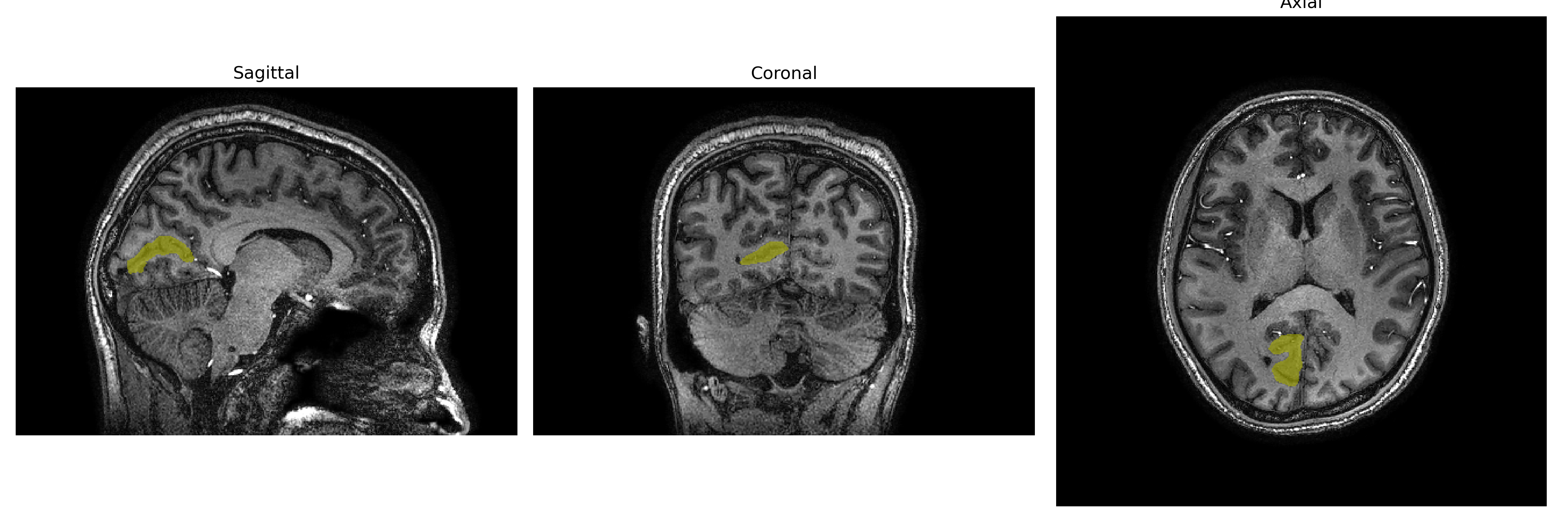
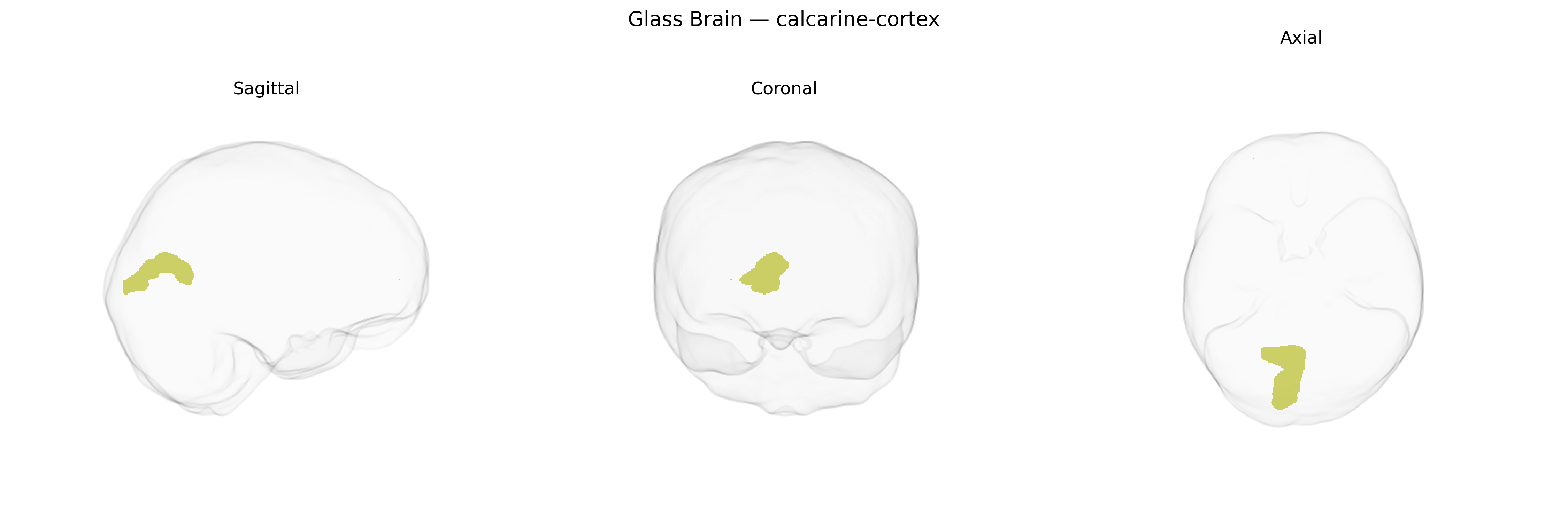
The right calcarine cortex, corresponding largely to the primary visual cortex (V1, Brodmann area 17) along the right calcarine sulcus of the occipital lobe, is a layered neocortical region specialized for the initial cortical processing of visual information. It receives highly organized retinotopic input from the lateral geniculate nucleus of the thalamus, with the upper and lower banks of the sulcus representing inferior and superior visual fields, respectively. Neurons in this region are tuned to fundamental visual features such as orientation, spatial frequency, contrast, and ocular dominance, forming columnar and laminar microcircuits that support edge detection and basic scene structure. Right-hemisphere calcarine cortex is especially important for processing visual information from the left visual hemifield and contributes to conscious visual perception, early visual integration, and the foundation of higher-order visual processing in extrastriate areas. Primary visual cortex
The right calcarine cortex, corresponding largely to primary visual cortex (V1) in the brainCOLOR atlas, has been implicated in multiple imaging-genetics and GWAS studies of brain structure and function, although locus- and gene-specific findings are typically reported at broader occipital or global cortical measures rather than calcarine-specific labels. Large-scale neuroimaging GWAS (e.g., ENIGMA, UK Biobank–based studies) have identified common variants near genes involved in neuronal development and synaptic function, such as CACHD1, HMGA2, KIAA0586, and others, associated with occipital cortical thickness or surface area measures that include the calcarine region, with some hemispheric asymmetry effects reported but not consistently restricted to the right side. Genetic correlations have been observed between occipital/calarine structural metrics and cognitive traits (educational attainment, intelligence), as well as with general brain size, suggesting partly shared polygenic influences. Functional and structural alterations of the calcarine cortex have been reported in genetic or strongly heritable conditions affecting visual processing and neurodevelopment (e.g., albinism, congenital blindness, certain monogenic visual pathway disorders), though these are typically described as downstream neuroanatomical consequences rather than loci identified in calcarine-specific GWAS. Additionally, imaging-genetics work in psychiatric and neurological disorders (schizophrenia, major depression, migraine, and some forms of epilepsy) has documented calcarine/occipital volume or activation differences linked to polygenic risk scores, but robust, replicated SNP-level associations that are uniquely and specifically assigned to the right calcarine cortex remain limited, with most evidence indicating a broadly polygenic, shared architecture with other visual and cortical regions.
Overview generated by GPT-4o (2026).
Region ID: 32
Hemisphere: Right
Atlas: brainCOLOR
Full Quality Version: Download MP4
Full Quality Version: Download MP4
Full Quality Version: Download MP4
Full Quality Version: Download MP4
Wali Sidiqyar*, Gaurav Rudravaram*, Elyssa M. McMaster, Trent M. Schwartz, Adam M. Saunders, Kurt G. Schilling, Bennett A. Landman "Introducing SPINS: A Shared Public Visualization Library of Neuroanatomical Structures." Medical Imaging with Deep Learning- short paper
This resource is licensed under CC0 1.0 Universal (Public Domain).