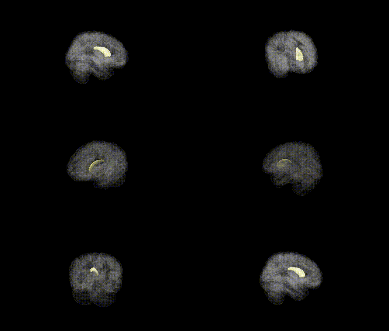
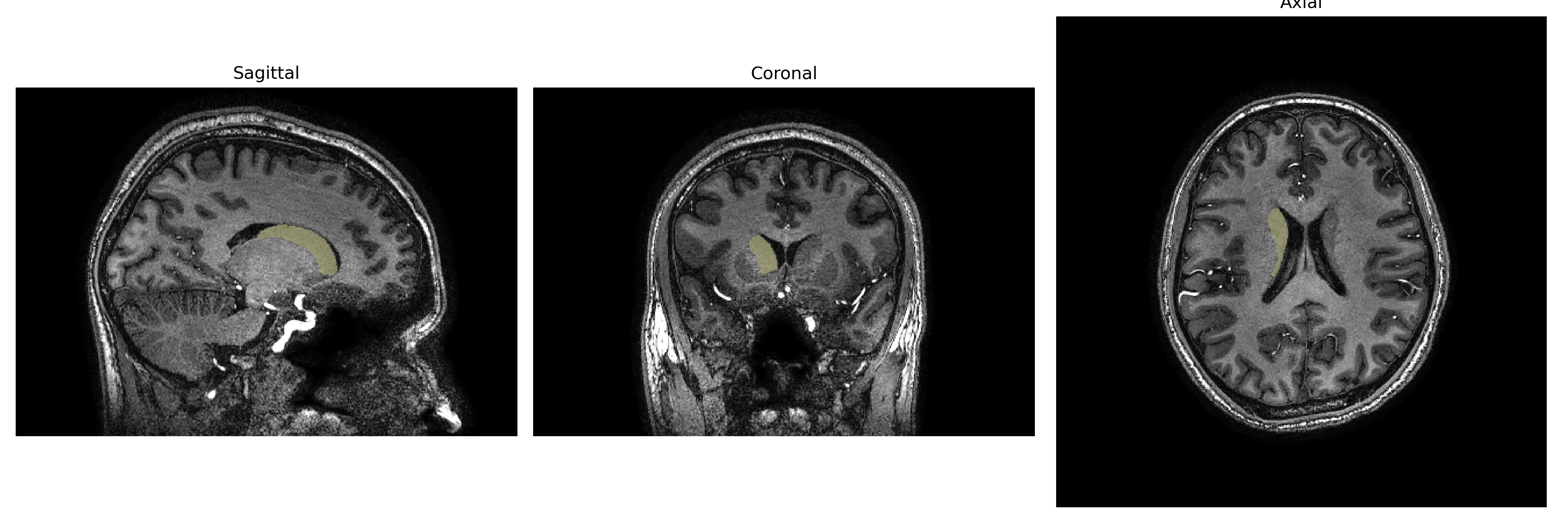
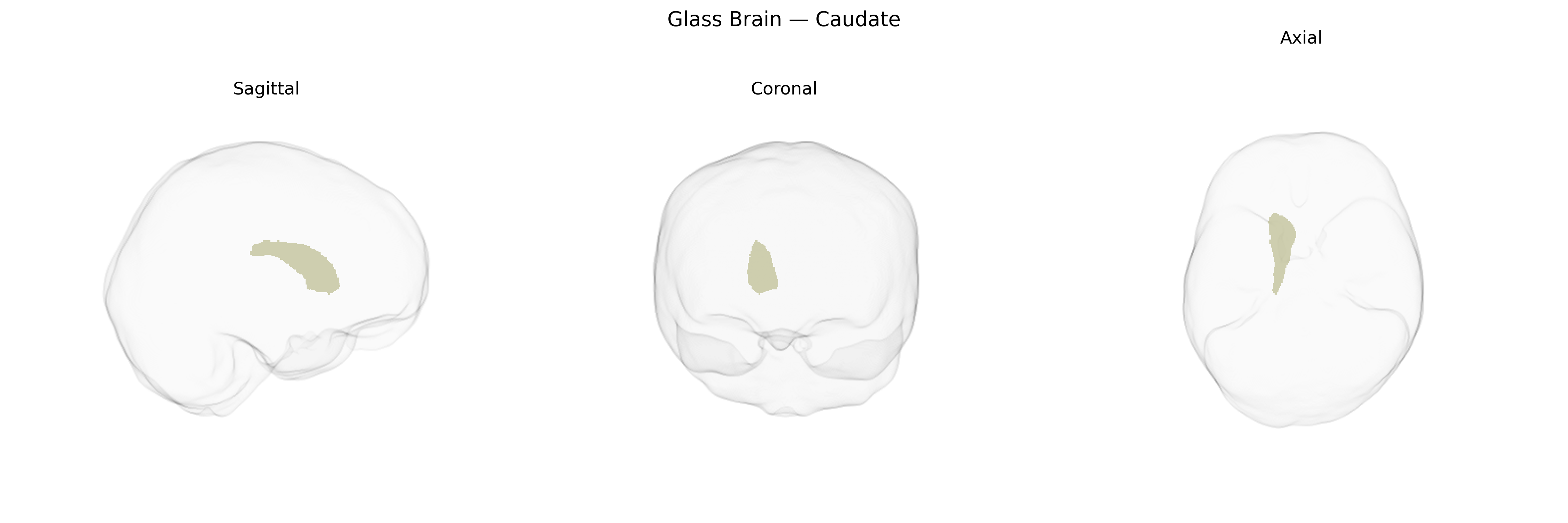
The Right Caudate, part of the caudate nucleus within the dorsal striatum of the basal ganglia, is a C-shaped gray matter structure located adjacent to the lateral ventricle in the right hemisphere. It plays a central role in motor control, associative learning, goal-directed behavior, and aspects of executive function, receiving dense glutamatergic inputs from frontal and parietal cortices and dopaminergic projections from the substantia nigra pars compacta. Its output is primarily GABAergic, projecting via the internal segment of the globus pallidus and substantia nigra pars reticulata to thalamic nuclei that modulate cortical activity. Functionally, the right caudate is implicated in action selection, habit formation, and cognitive flexibility, and structural or functional abnormalities in this region have been associated with disorders such as Parkinson’s disease, Huntington’s disease, obsessive–compulsive disorder, and attention-deficit/hyperactivity disorder. Caudate nucleus
The right caudate, as defined in the brainCOLOR Atlas, has been implicated in multiple genetic associations through GWAS and imaging-genetics studies, particularly those examining subcortical volumes and neuropsychiatric risk. Large-scale consortia such as ENIGMA and UK Biobank–based studies have identified common variants near or within genes including FAT3, DCC, PTCH1, HMGA2, and others that influence caudate volume bilaterally, with some loci showing hemisphere-specific or asymmetric effects that encompass the right caudate. Polygenic risk for schizophrenia, ADHD, and major depressive disorder has been associated with structural alterations in caudate volume, and risk variants in dopaminergic, glutamatergic, and synaptic genes (for example, DRD2-related loci and CACNA1C-related pathways) have been linked to caudate morphology or function in case–control or endophenotype designs. GWAS of cognitive performance, educational attainment, and general intelligence have also shown that polygenic scores for these traits correlate with right caudate volume, supporting a genetically mediated role in executive functions and learning. Additionally, variants associated with Parkinson’s disease and other movement disorders, including loci related to dopaminergic neurodegeneration, have been connected to caudate structural and functional differences, although most findings are not strictly right-lateralized. Overall, genetic studies converge on the right caudate as a heritable subcortical structure whose volume and connectivity are shaped by multiple common variants and polygenic architectures that overlap with risk for psychiatric, cognitive, and movement-related phenotypes.
Overview generated by GPT-4o (2026).
Region ID: 5
Hemisphere: Right
Atlas: brainCOLOR
Full Quality Version: Download MP4
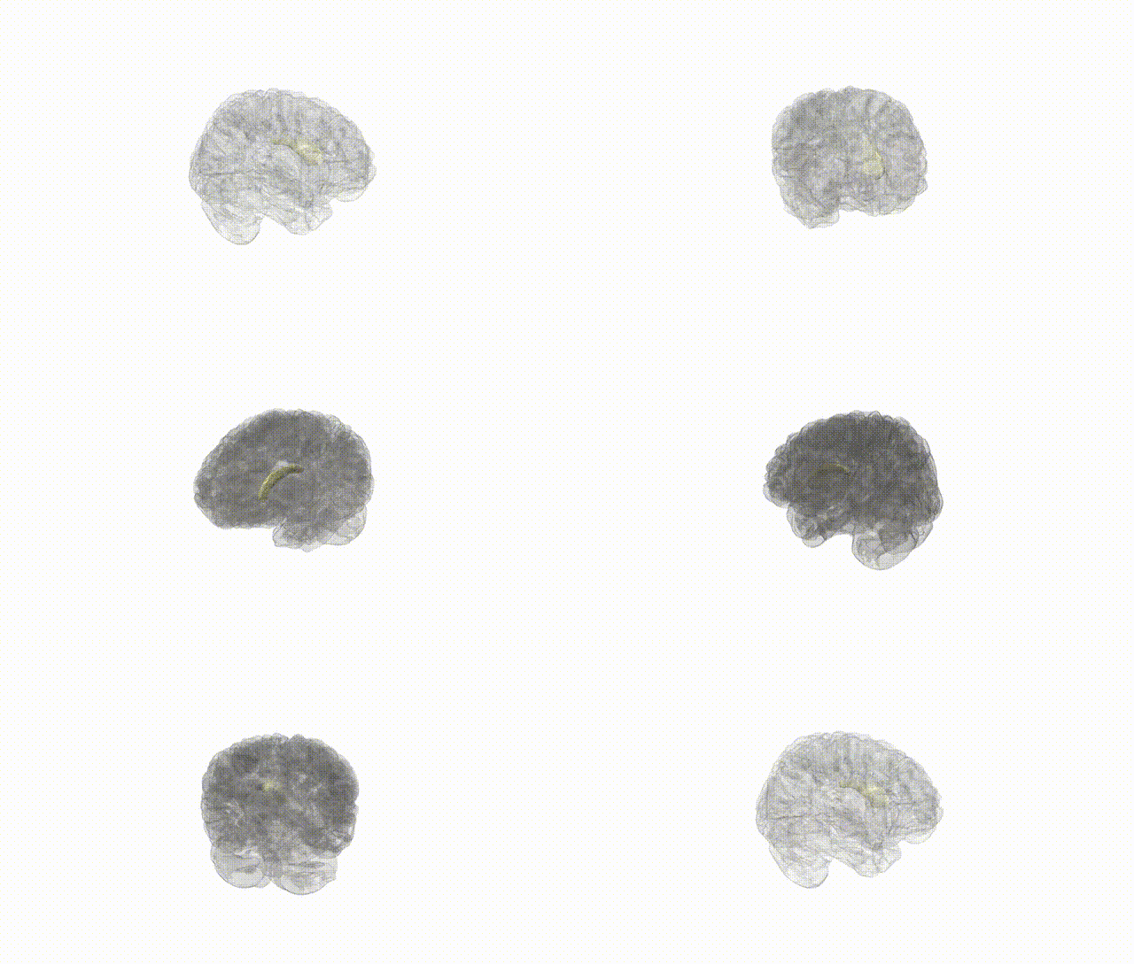
Full Quality Version: Download MP4
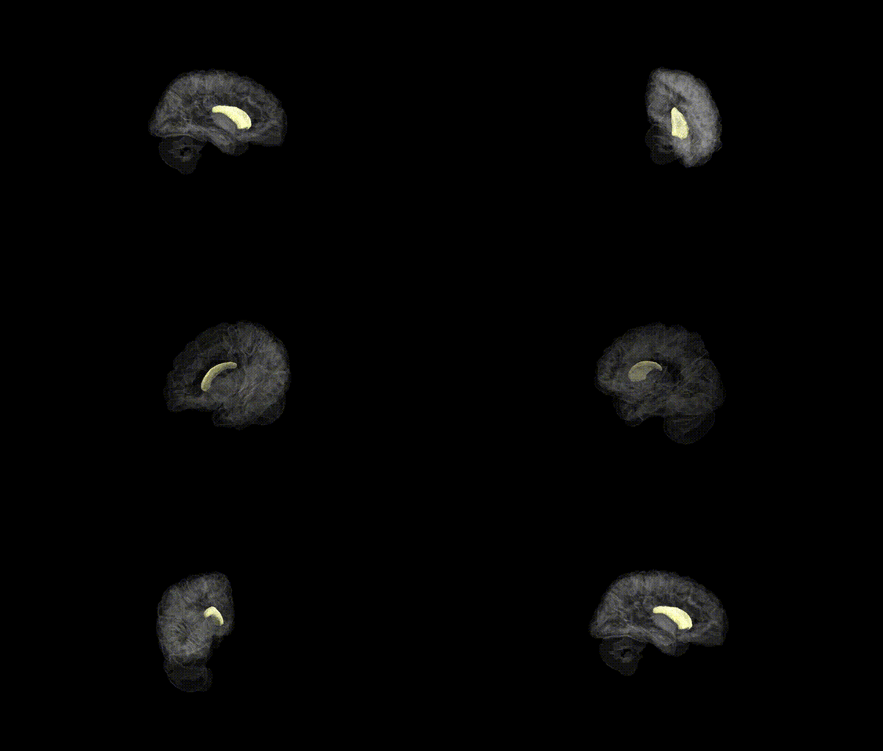
Full Quality Version: Download MP4
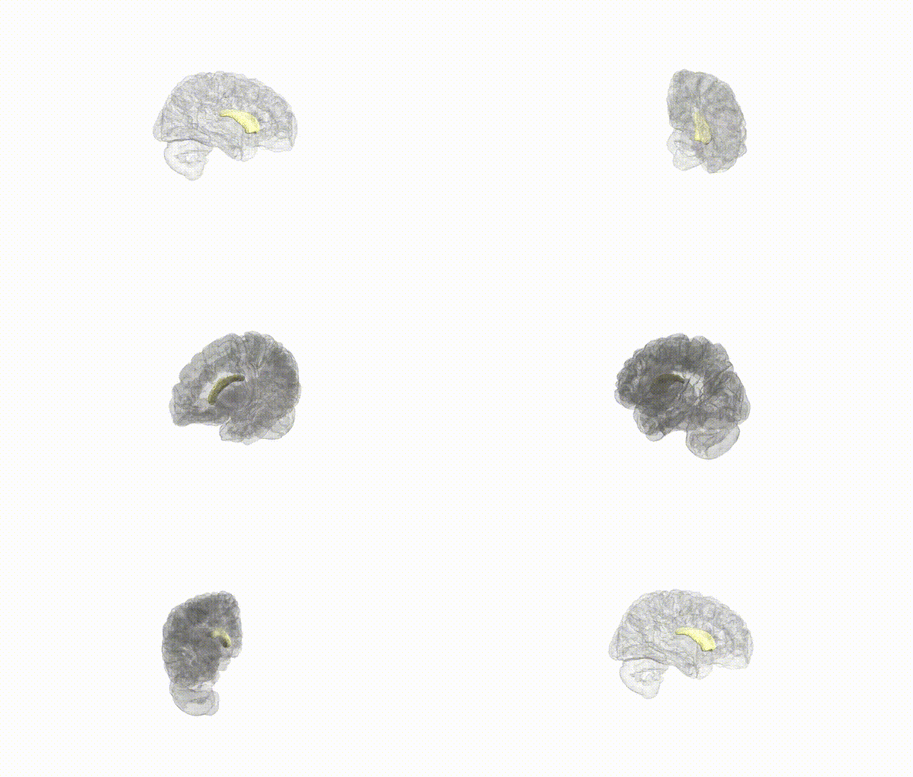
Full Quality Version: Download MP4
Wali Sidiqyar*, Gaurav Rudravaram*, Elyssa M. McMaster, Trent M. Schwartz, Adam M. Saunders, Kurt G. Schilling, Bennett A. Landman "Introducing SPINS: A Shared Public Visualization Library of Neuroanatomical Structures." Medical Imaging with Deep Learning- short paper
This resource is licensed under CC0 1.0 Universal (Public Domain).