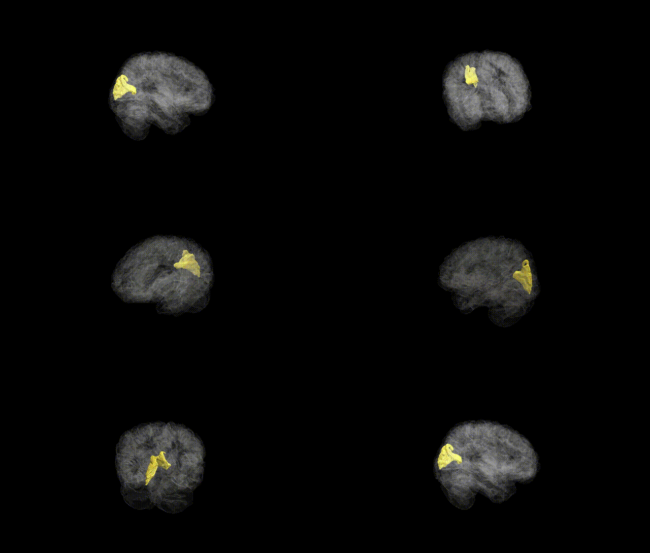
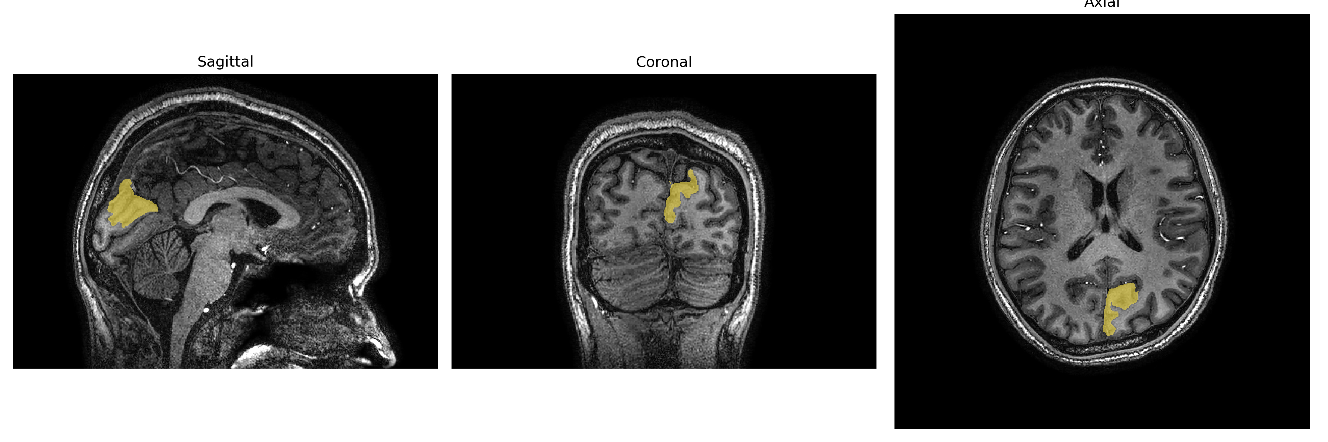
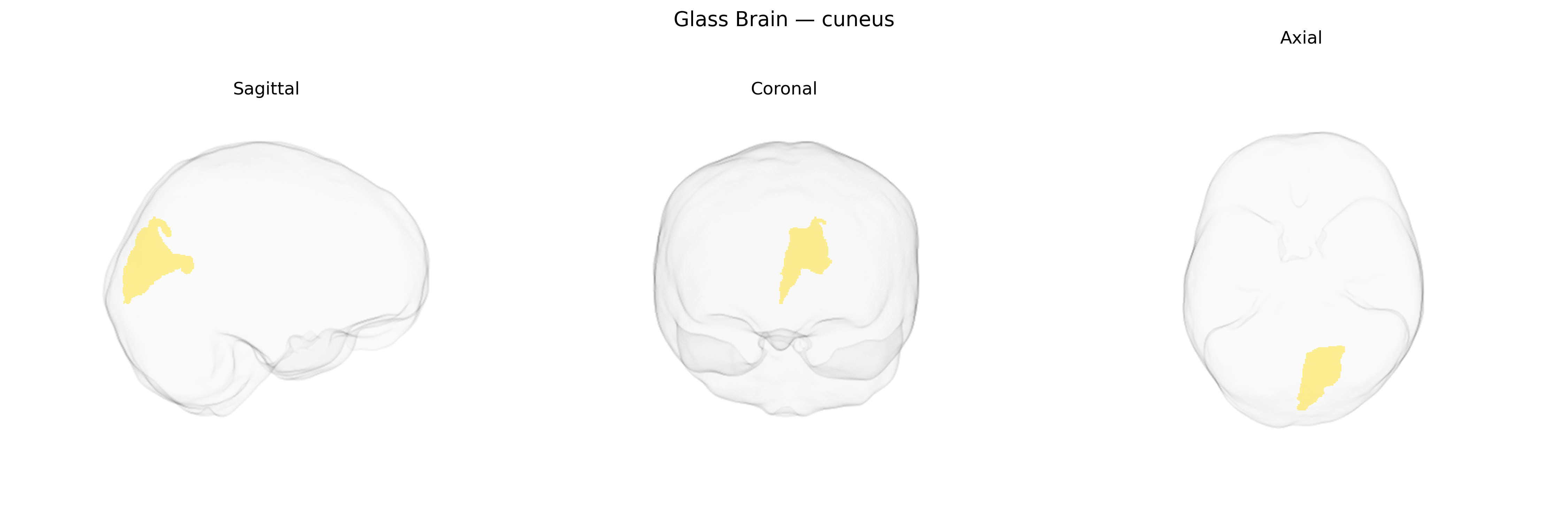
The left cuneus is a wedge-shaped cortical region of the occipital lobe situated between the parieto-occipital sulcus and the calcarine sulcus on the medial surface of the hemisphere. It forms part of the primary and secondary visual cortical areas and is involved in basic visual processing, including pattern, motion, and visual field representation, with a particular role in processing information from the lower visual field. The left cuneus participates in higher-order visuospatial integration and visual attention, and its activity has been implicated in tasks involving visual imagery, orientation, and eye movements. As delineated in parcellation schemes such as the brainCOLOR Atlas, the left cuneus is defined by its cytoarchitectonic features and connectivity with other visual and parietal association areas. Cuneus
The left cuneus, a primary and associative visual cortex region in the occipital lobe, has been implicated in multiple genome-wide association studies, though typically as part of broader occipital or visual network phenotypes rather than as an isolated structure. Large-scale imaging genetics consortia (e.g., ENIGMA, UK Biobank) have identified common variants near genes involved in neurodevelopment, synaptic function, and axonal guidance (such as those in glutamatergic and calcium signaling pathways) that associate with cuneus cortical thickness, surface area, or functional connectivity, though specific brainCOLOR Atlas–labeled “Left cuneus” GWAS hits are not yet standardly reported as standalone loci. Polygenic risk for schizophrenia, major depressive disorder, bipolar disorder, and autism spectrum disorder has been associated with structural and functional alteration in occipital and cuneus regions, including reduced cortical thickness or altered resting-state connectivity, suggesting that shared genetic architectures influencing visual and higher-order cognitive networks can affect this region. In addition, variants linked to cognitive traits (general intelligence, educational attainment), visual processing performance, and migraine have been associated with differential activation or morphology in cuneus/occipital areas, and several Alzheimer’s disease and general brain aging GWAS highlight occipital cortical measures that include the cuneus, indicating that genetic risk for neurodegeneration may partly manifest in this region’s structure and connectivity.
Overview generated by GPT-4o (2026).
Region ID: 37
Hemisphere: Left
Atlas: brainCOLOR
Full Quality Version: Download MP4
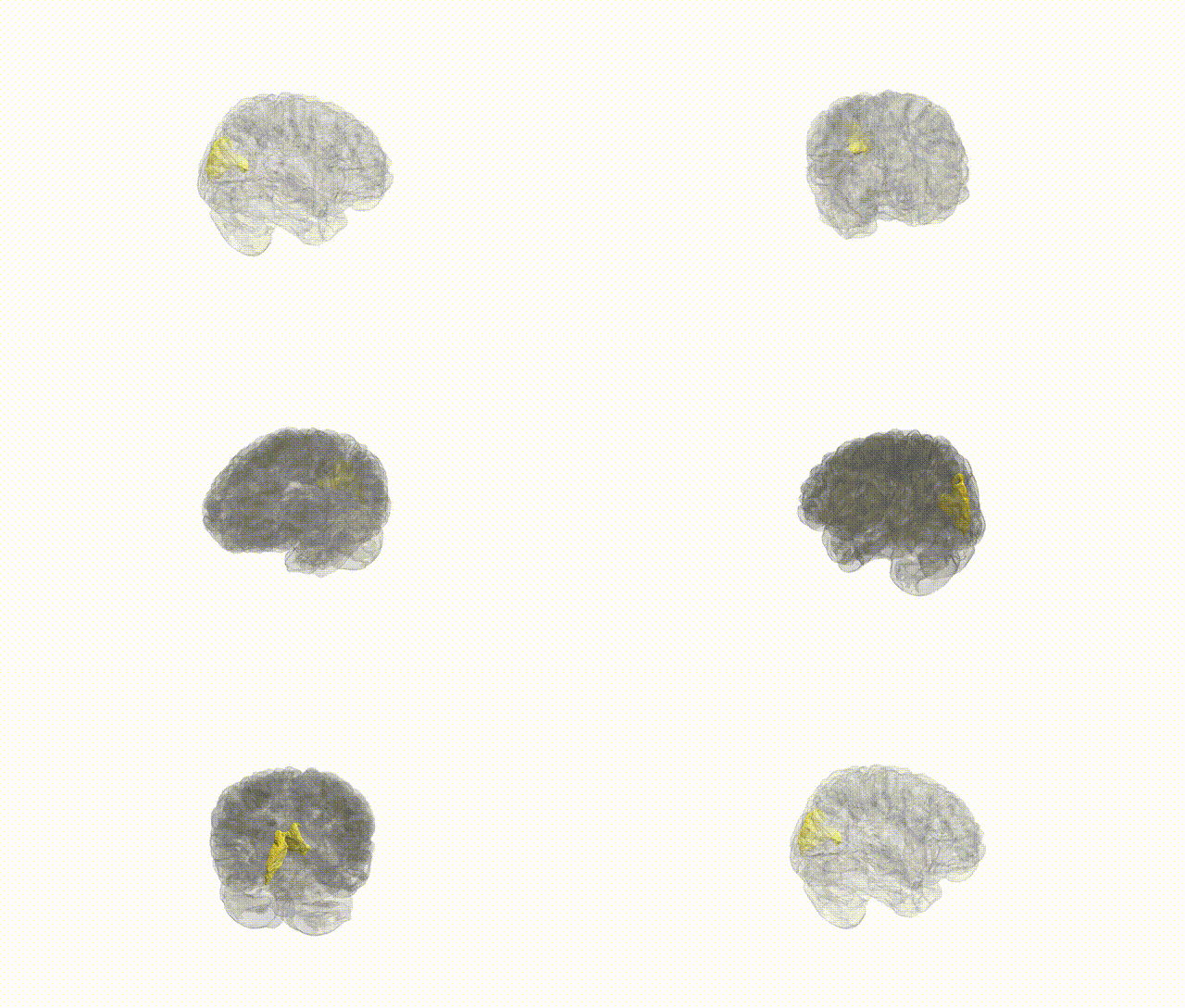
Full Quality Version: Download MP4
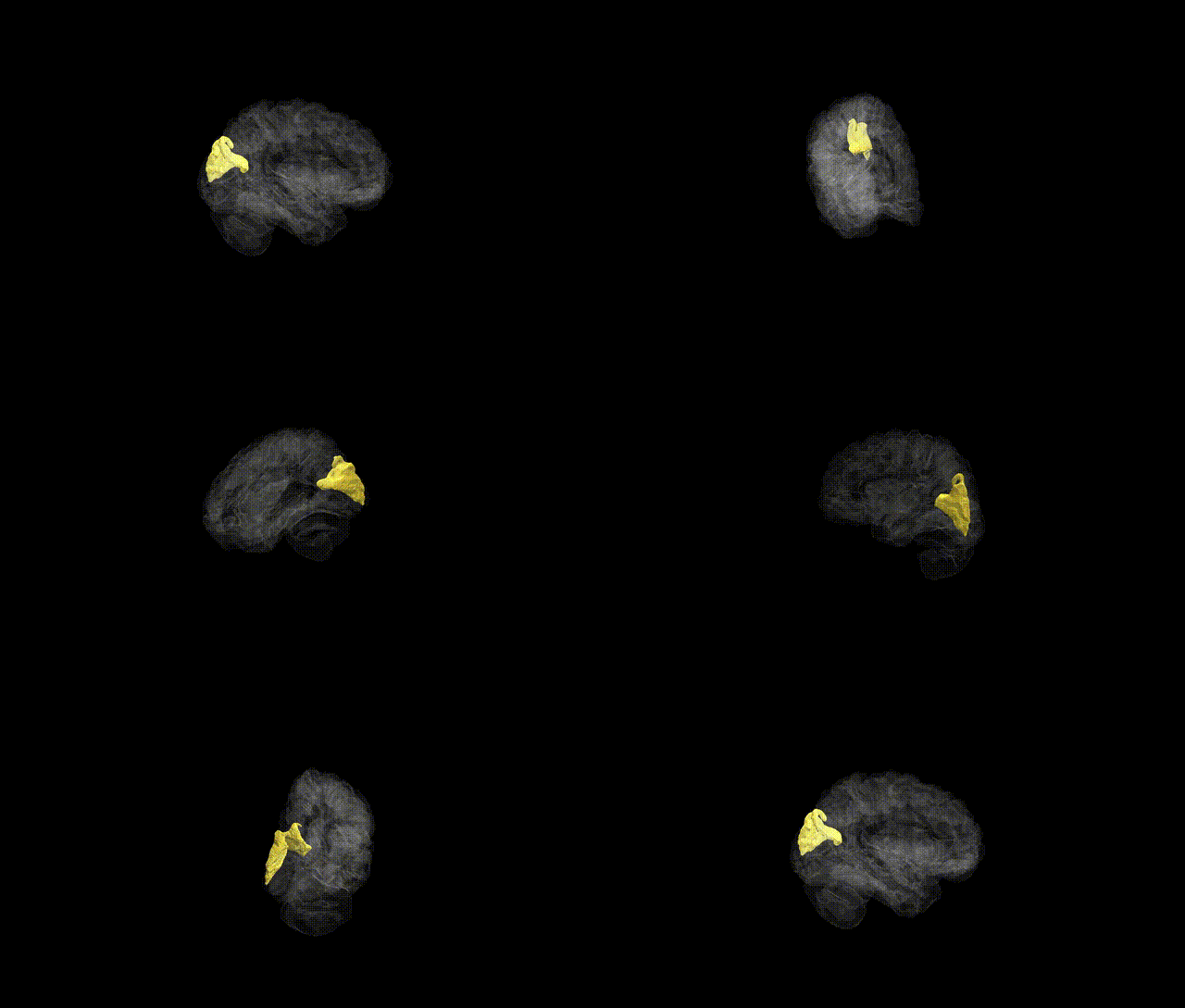
Full Quality Version: Download MP4
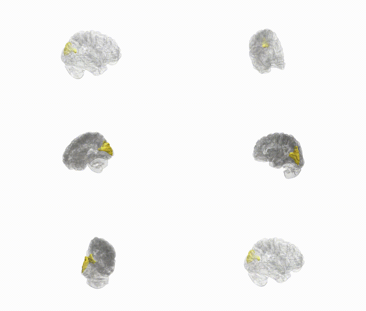
Full Quality Version: Download MP4
Wali Sidiqyar*, Gaurav Rudravaram*, Elyssa M. McMaster, Trent M. Schwartz, Adam M. Saunders, Kurt G. Schilling, Bennett A. Landman "Introducing SPINS: A Shared Public Visualization Library of Neuroanatomical Structures." Medical Imaging with Deep Learning- short paper
This resource is licensed under CC0 1.0 Universal (Public Domain).