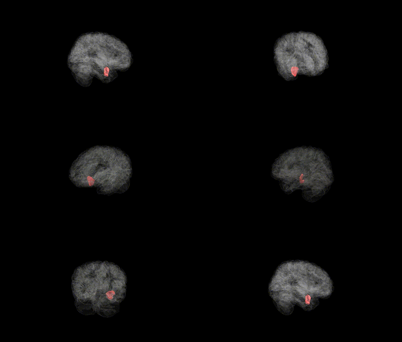
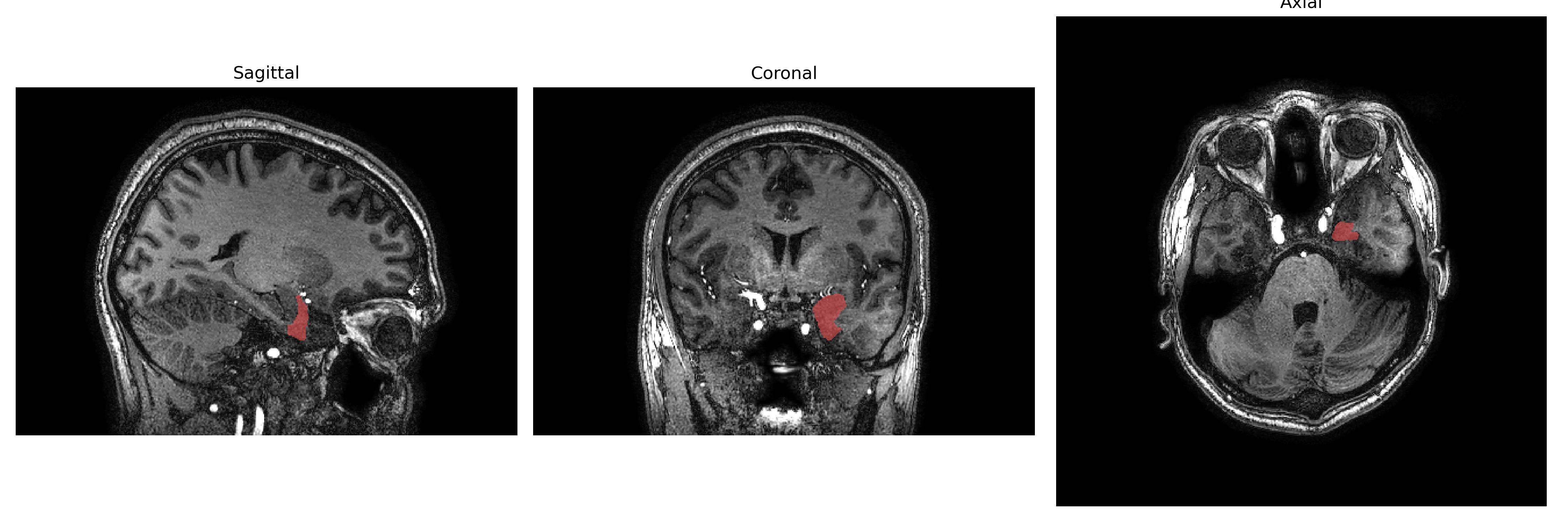
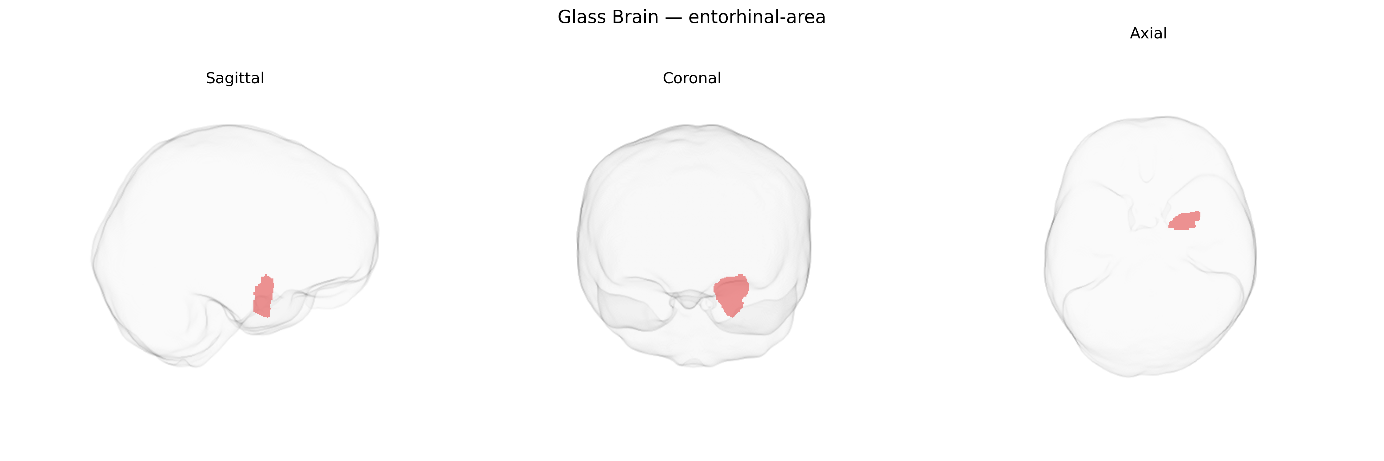
The left entorhinal area is a medial temporal lobe cortical region that forms a critical interface between the neocortex and the hippocampal formation, serving as a major input–output gateway for declarative memory and spatial navigation networks. It receives highly processed multimodal sensory information from widespread association cortices and projects to the hippocampus via the perforant path, while also participating in reciprocal circuits with the subiculum and parahippocampal regions. Neurons in this area, including grid cells and other spatially tuned cell types, contribute to the encoding of spatial context and episodic memory representations. The entorhinal area is among the earliest sites of pathological change in Alzheimer’s disease and other neurodegenerative conditions, where neurofibrillary tangles and cell loss can disrupt memory formation and navigation. The left hemisphere localization reflects lateralization patterns often associated with verbal and language-related aspects of episodic memory. Entorhinal cortex
Genetic associations specifically targeting the Left entorhinal area as defined in the brainCOLOR atlas are limited, but broader imaging-genetics and GWAS work on entorhinal cortex structure and function implicates several loci and pathways. Variants in and near APOE, particularly ε4, show strong associations with entorhinal cortical thinning, atrophy, and altered functional connectivity, consistent with the region’s early involvement in Alzheimer’s disease (AD) pathology. Large MRI-based GWAS of cortical thickness and surface area have identified common variants in genes related to lipid metabolism, neuronal development, and synaptic function (for example, loci near WNT3, KIAA0586, and other neurodevelopmental and axon-guidance genes) that influence medial temporal and entorhinal morphometry, though effects are often reported bilaterally or for broader medial temporal lobe clusters rather than a left-lateralized parcel. Polygenic risk scores for AD and, to a lesser degree, for schizophrenia and major depressive disorder have been associated with reduced entorhinal volume or cortical thickness in population cohorts, suggesting a shared genetic architecture between psychiatric and neurodegenerative liability and entorhinal structure. In addition, risk variants for primary tauopathies (such as MAPT haplotypes) and genes involved in amyloid and tau processing show enrichment of effects on entorhinal and adjacent medial temporal measures, aligning with this region’s role in episodic memory and vulnerability to neurofibrillary tangle accumulation, although highly specific left entorhinal–only genetic signals remain sparsely characterized.
Overview generated by GPT-4o (2026).
Region ID: 39
Hemisphere: Left
Atlas: brainCOLOR
Full Quality Version: Download MP4
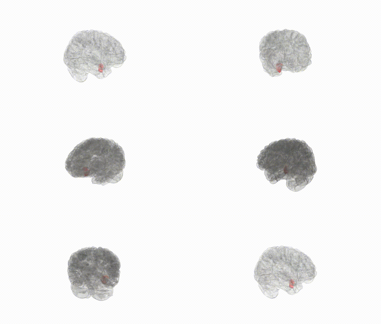
Full Quality Version: Download MP4
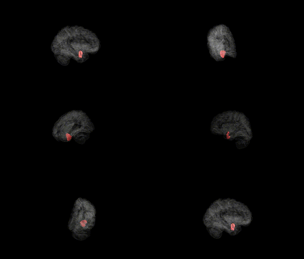
Full Quality Version: Download MP4
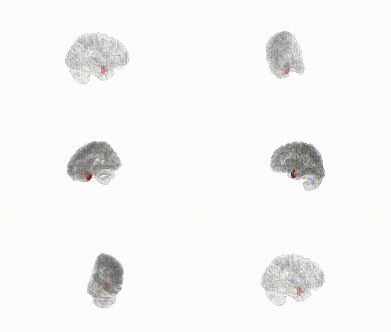
Full Quality Version: Download MP4
Wali Sidiqyar*, Gaurav Rudravaram*, Elyssa M. McMaster, Trent M. Schwartz, Adam M. Saunders, Kurt G. Schilling, Bennett A. Landman "Introducing SPINS: A Shared Public Visualization Library of Neuroanatomical Structures." Medical Imaging with Deep Learning- short paper
This resource is licensed under CC0 1.0 Universal (Public Domain).