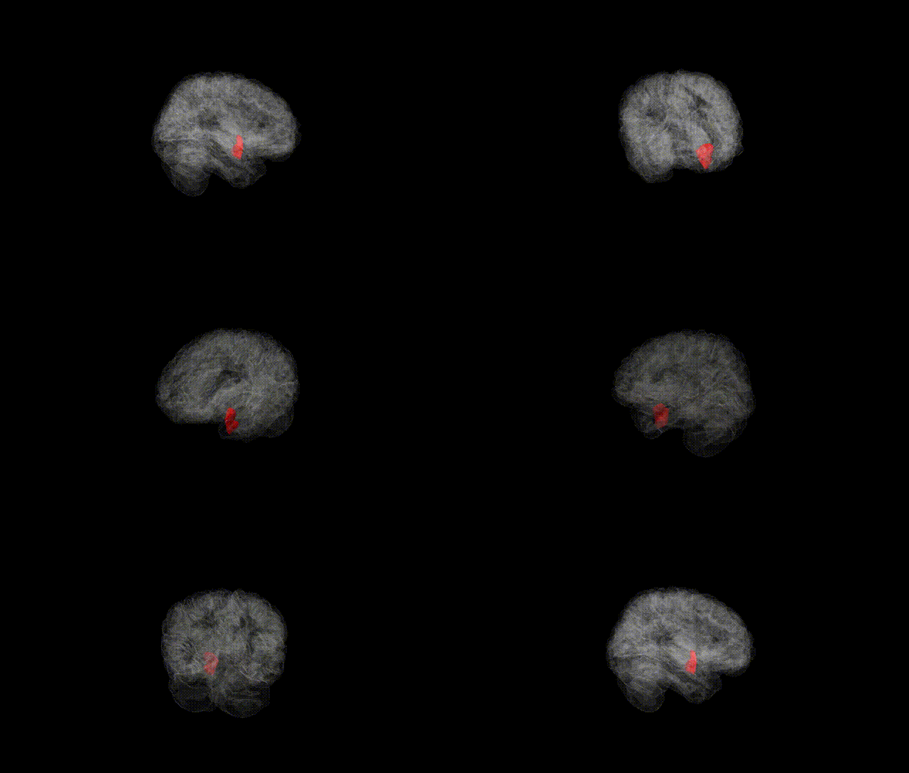
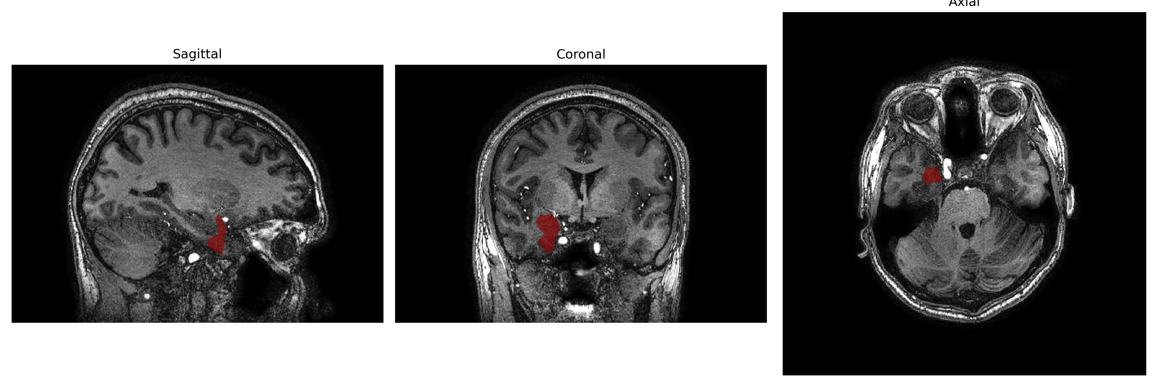
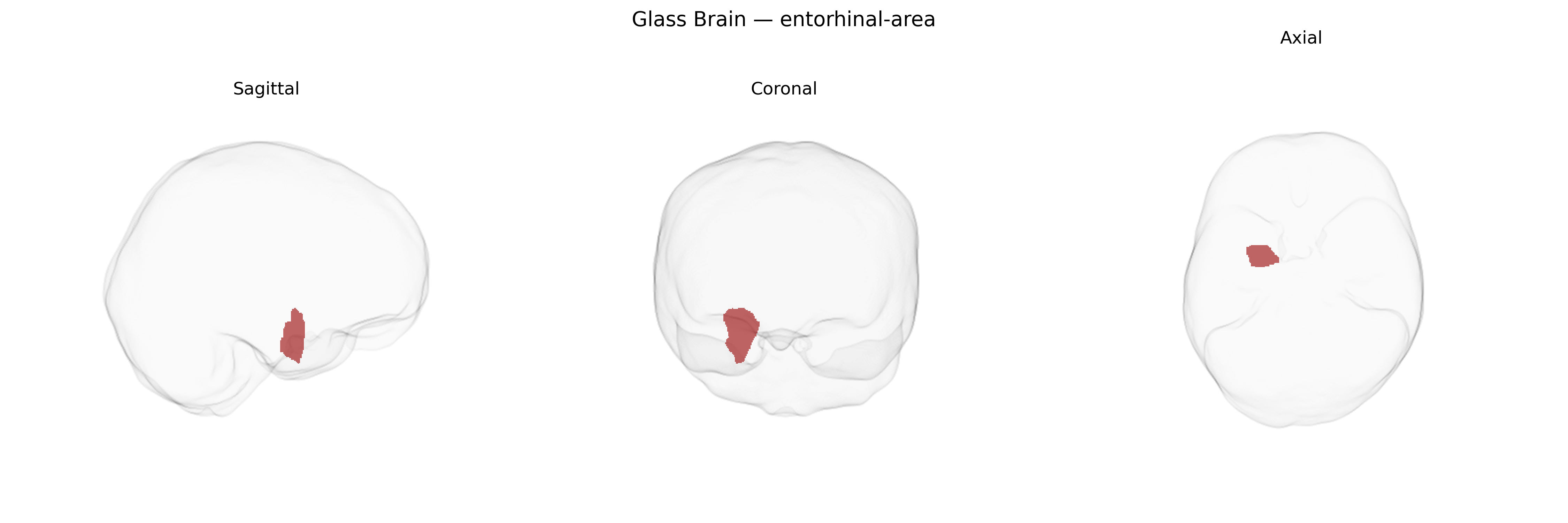
The right entorhinal-area is a cortical region of the medial temporal lobe that forms a critical interface between widespread neocortical areas and the hippocampal formation, particularly the dentate gyrus and CA fields. It receives highly processed multimodal sensory and associative input and conveys this information to the hippocampus via the perforant path, playing a central role in episodic memory formation, spatial navigation, and contextual representation. Cytoarchitectonically, it is characterized by distinct lamination patterns and specialized neuronal populations, including grid cells and other spatially tuned neurons that support map-like coding of the environment. The right entorhinal-area often shows early pathological changes in Alzheimer’s disease, including tau pathology and neurofibrillary tangles, making it a key focus in neurodegenerative research. No direct article exists; see the related structure: Entorhinal cortex.
The right entorhinal cortex, as defined in parcellations such as the brainCOLOR atlas, has been implicated in multiple genetic and GWAS-based findings, primarily through studies of hippocampal–medial temporal lobe structure and neurodegeneration. Twin and SNP-heritability analyses show that entorhinal cortical thickness and volume are moderately to highly heritable, with common variants explaining a substantial fraction of inter-individual variability. Large neuroimaging GWAS (e.g., ENIGMA, UK Biobank) have identified associations between entorhinal measures and variants in or near genes involved in neurodevelopment, synaptic organization, and neurodegenerative pathways, including loci overlapping or proximal to APOE (particularly ε4), BIN1, and other Alzheimer’s disease risk genes, consistent with the entorhinal cortex being one of the earliest sites of tau pathology. Genetic risk scores for Alzheimer’s disease and related dementias show robust associations with reduced entorhinal thickness or volume, and variants influencing general cognitive ability and memory performance sometimes demonstrate parallel effects on entorhinal structure. Additional GWAS of brain aging and cortical atrophy patterns indicate that polygenic architectures contributing to age-related cortical thinning, especially in medial temporal regions, also affect the right entorhinal area, linking it genetically to trajectories of memory decline, mild cognitive impairment, and Alzheimer’s disease progression.
Overview generated by GPT-4o (2026).
Region ID: 38
Hemisphere: Right
Atlas: brainCOLOR
Full Quality Version: Download MP4
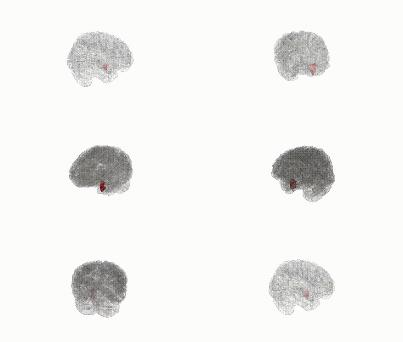
Full Quality Version: Download MP4
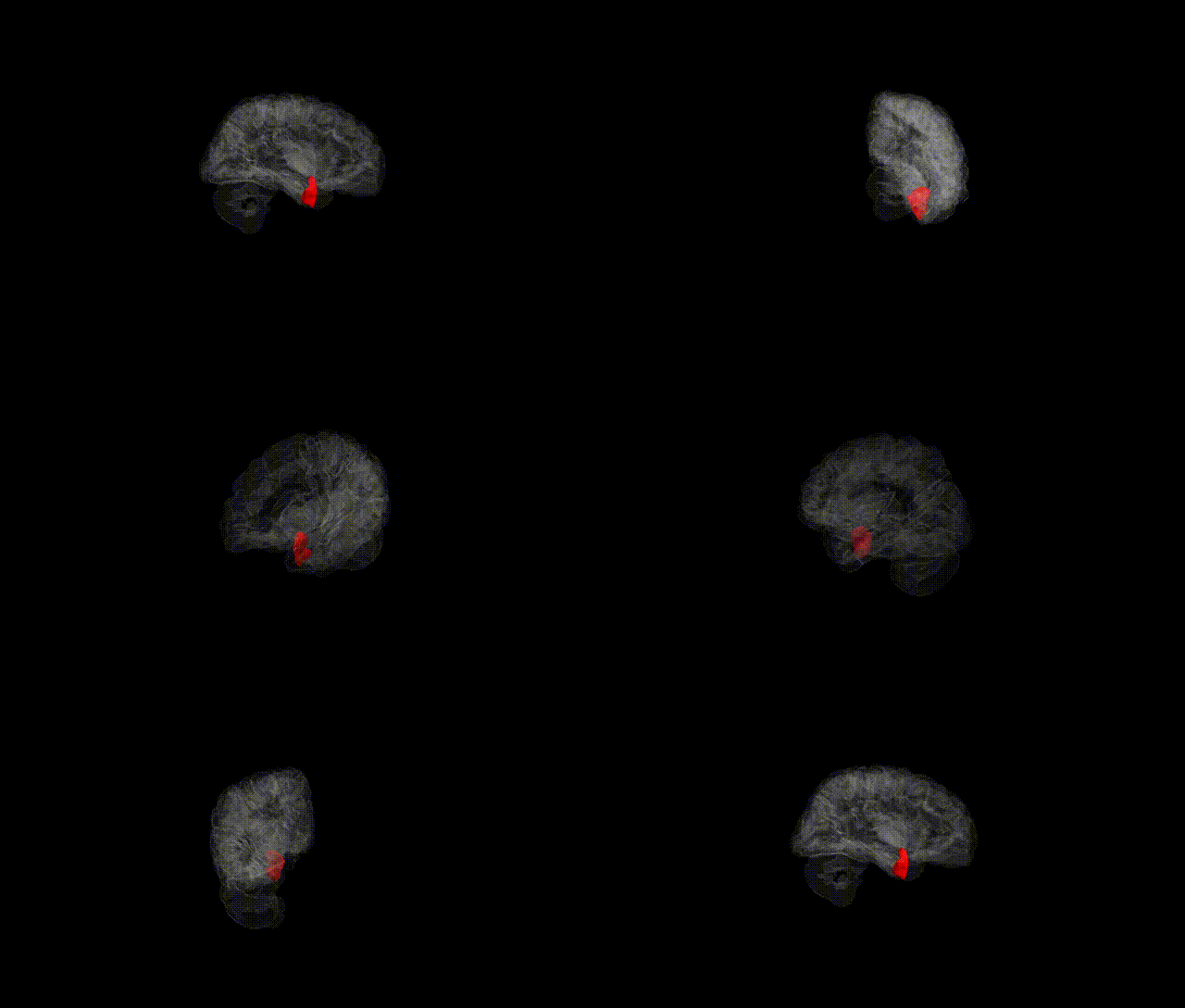
Full Quality Version: Download MP4
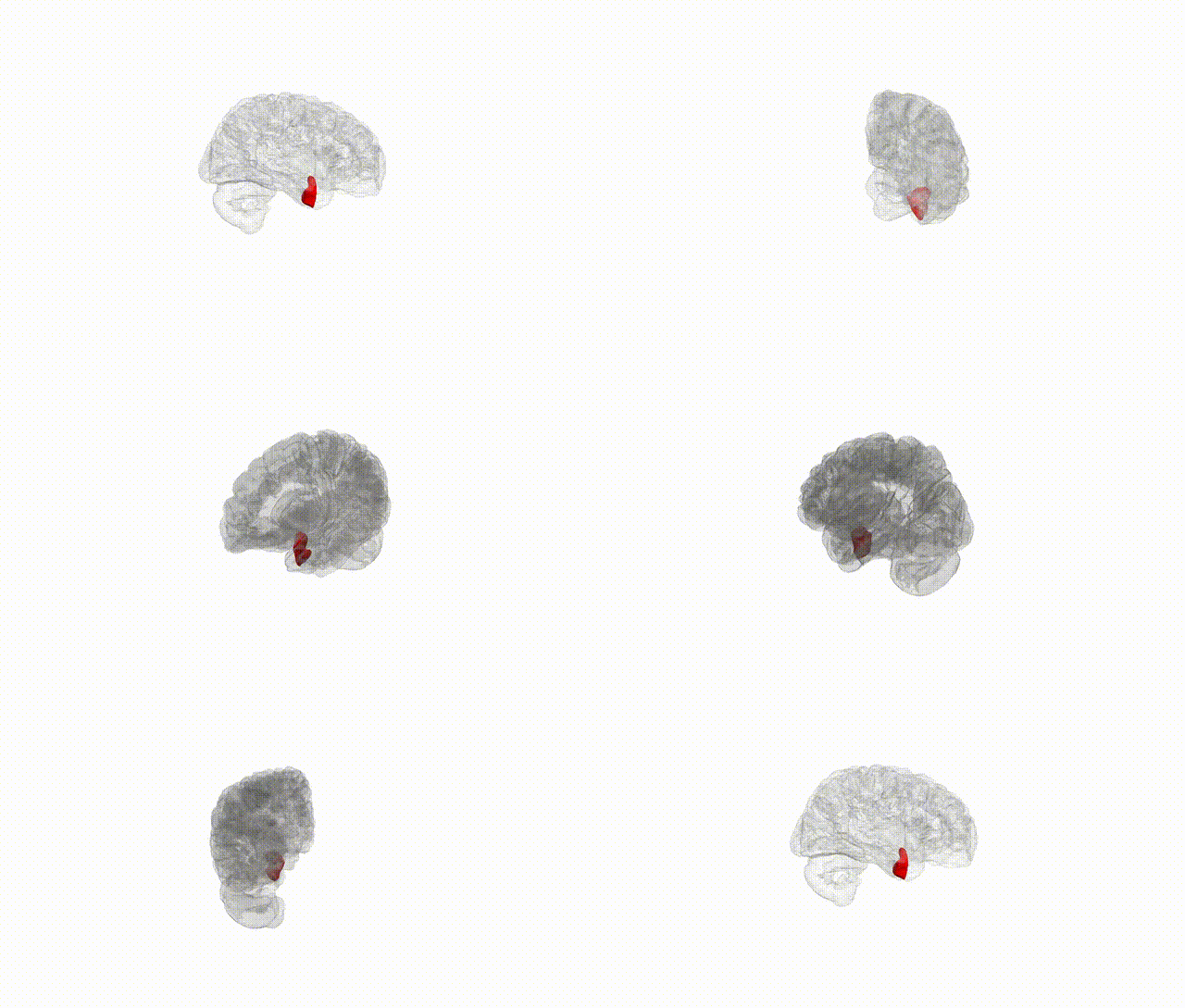
Full Quality Version: Download MP4
Wali Sidiqyar*, Gaurav Rudravaram*, Elyssa M. McMaster, Trent M. Schwartz, Adam M. Saunders, Kurt G. Schilling, Bennett A. Landman "Introducing SPINS: A Shared Public Visualization Library of Neuroanatomical Structures." Medical Imaging with Deep Learning- short paper
This resource is licensed under CC0 1.0 Universal (Public Domain).