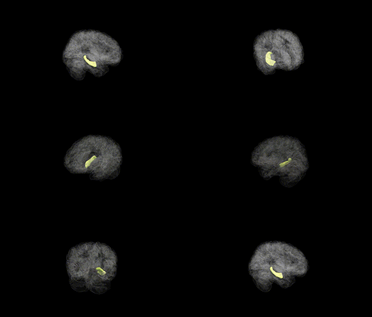
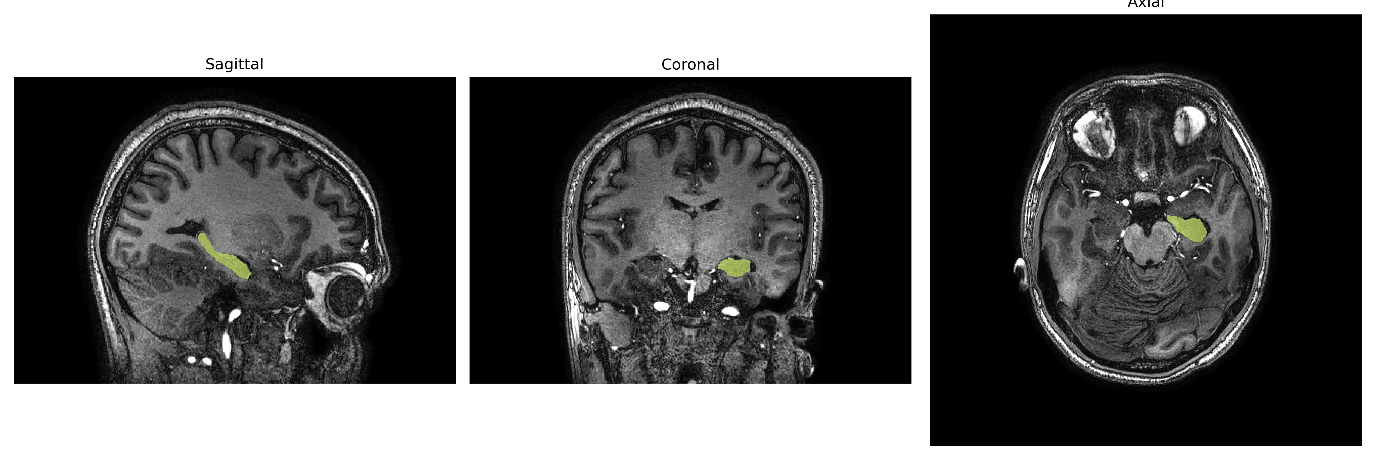
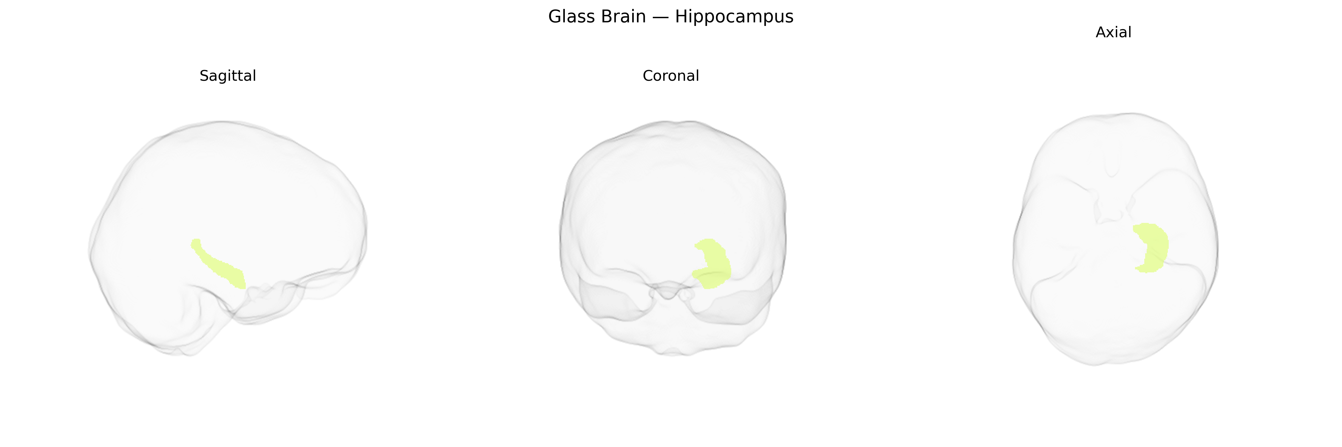
The Left Hippocampus is a medial temporal lobe structure that forms part of the hippocampal formation and plays a central role in declarative memory, spatial navigation, and the consolidation of information from short-term to long-term storage. It is anatomically organized along a longitudinal axis and comprises subfields including CA1–CA4, dentate gyrus, and subiculum, with dense reciprocal connections to the entorhinal cortex, amygdala, prefrontal cortex, and other limbic and association areas. Functionally, the left hippocampus is particularly implicated in verbal and narrative memory processes, contextual encoding, and episodic recollection, often showing lateralized specialization relative to the right hippocampus. It is highly vulnerable to hypoxic-ischemic injury, neurodegenerative processes such as Alzheimer’s disease, and mesial temporal lobe epilepsy, where volume loss and sclerosis are common. Histologically, it is characterized by layered allocortical organization, prominent pyramidal cell populations, and plastic synaptic circuits that support long-term potentiation and other mechanisms underlying learning and memory. Left Hippocampus
The left hippocampus, as defined in the brainCOLOR atlas, shows robust heritability and has been implicated in numerous genome-wide association studies (GWAS) of subcortical brain volumes, episodic memory, and neuropsychiatric and neurodegenerative disorders. Large-scale imaging genetics consortia (e.g., ENIGMA, UK Biobank) have identified common variants in and around genes such as HMGA2, MSRB3, DPP4, ASTN2, SLC4A10, and loci near APOE and MAPT as associated with hippocampal volume, with some effects showing lateralization or region-specific patterns that include the left hippocampus. Reduced left hippocampal volume and altered connectivity have been genetically linked to increased risk for Alzheimer’s disease (especially in carriers of APOE ε4), schizophrenia, major depressive disorder, bipolar disorder, and post-traumatic stress disorder, and polygenic risk scores for these conditions often correlate with left hippocampal structural differences. Variants in genes involved in neurodevelopment, synaptic plasticity, and neuroinflammation (for example in BDNF pathways, complement system genes, and microglial function genes) have also been associated with hippocampal morphology and memory performance. Additionally, GWAS of cognitive traits such as general intelligence, educational attainment, and memory show partial genetic overlap with loci influencing left hippocampal volume, suggesting a shared polygenic architecture between hippocampal structure, cognitive function, and vulnerability to psychiatric and neurodegenerative disease.
Overview generated by GPT-4o (2026).
Region ID: 10
Hemisphere: Left
Atlas: brainCOLOR
Full Quality Version: Download MP4
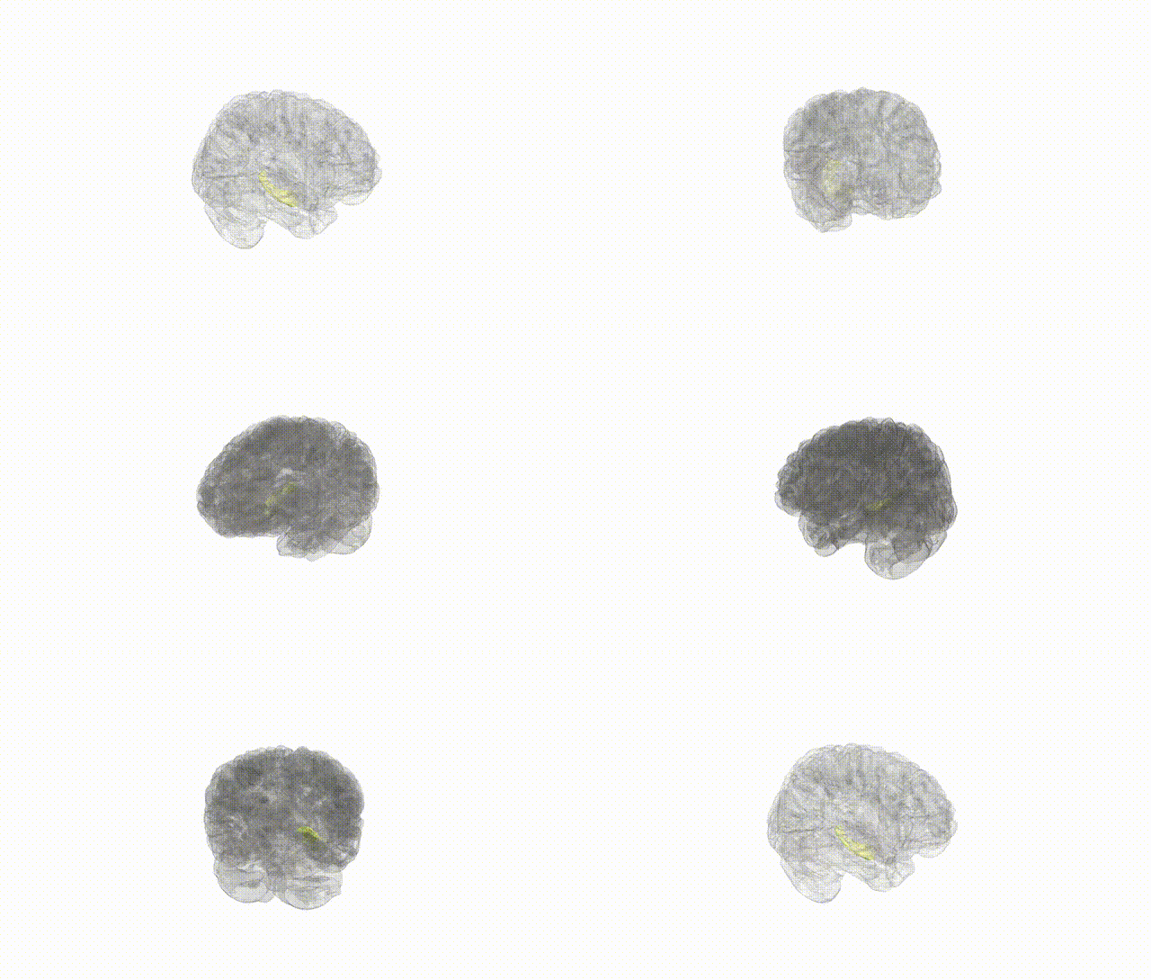
Full Quality Version: Download MP4
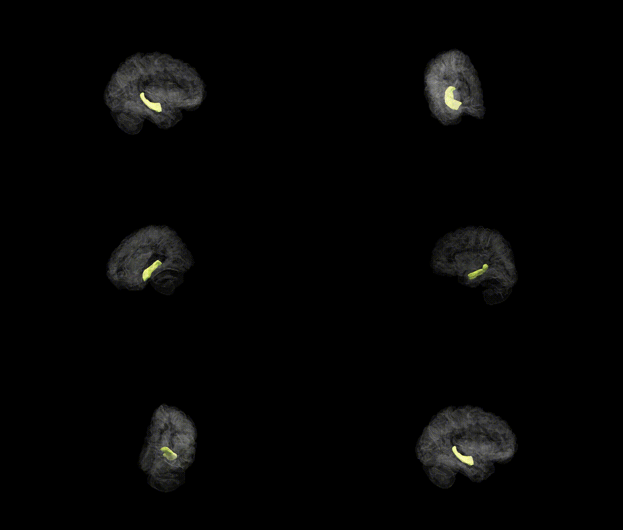
Full Quality Version: Download MP4
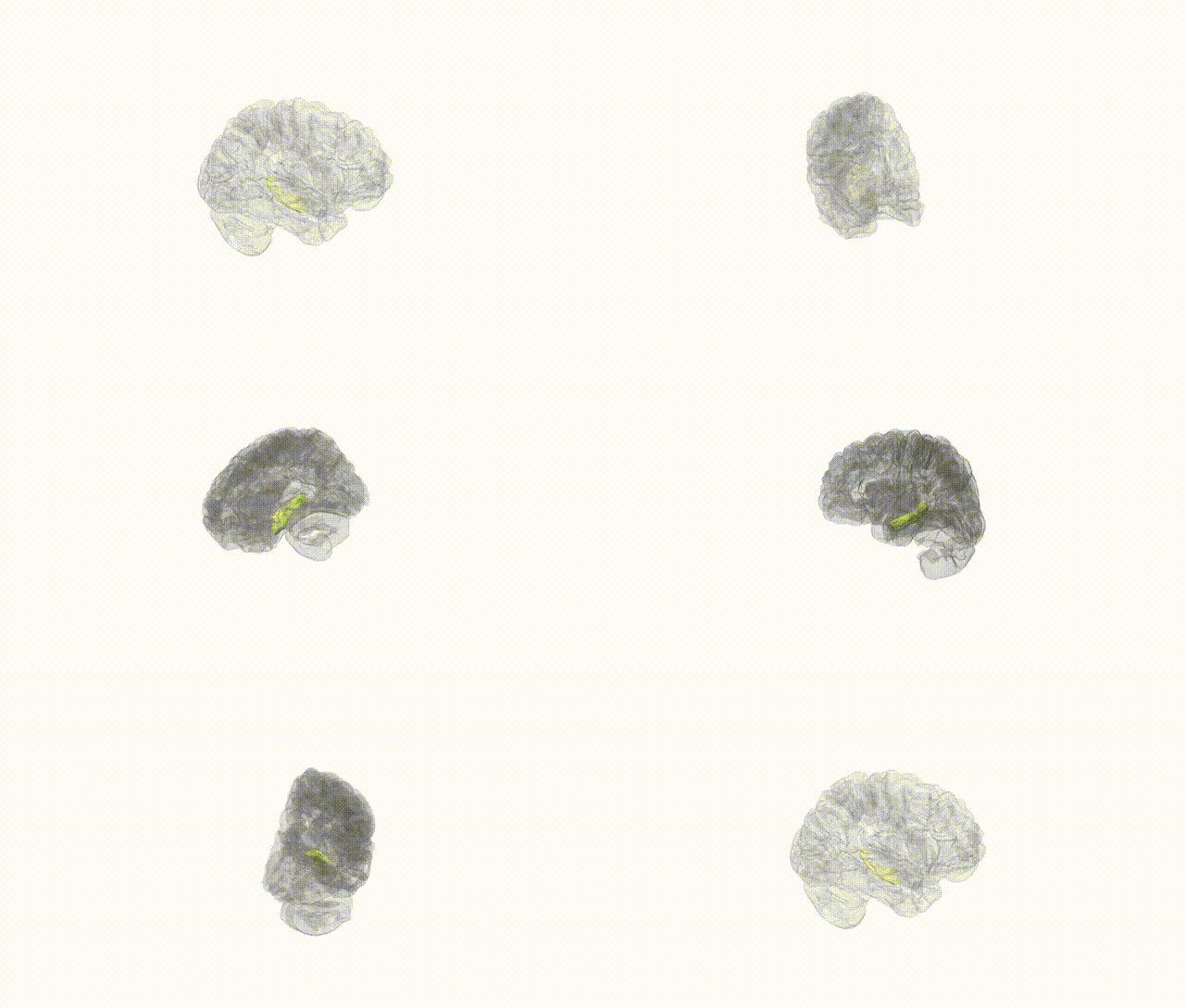
Full Quality Version: Download MP4
Wali Sidiqyar*, Gaurav Rudravaram*, Elyssa M. McMaster, Trent M. Schwartz, Adam M. Saunders, Kurt G. Schilling, Bennett A. Landman "Introducing SPINS: A Shared Public Visualization Library of Neuroanatomical Structures." Medical Imaging with Deep Learning- short paper
This resource is licensed under CC0 1.0 Universal (Public Domain).