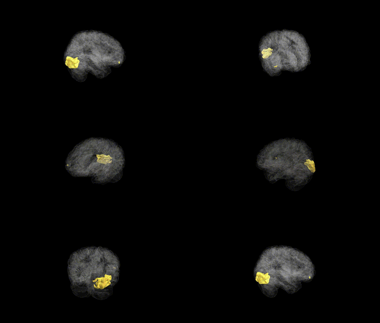
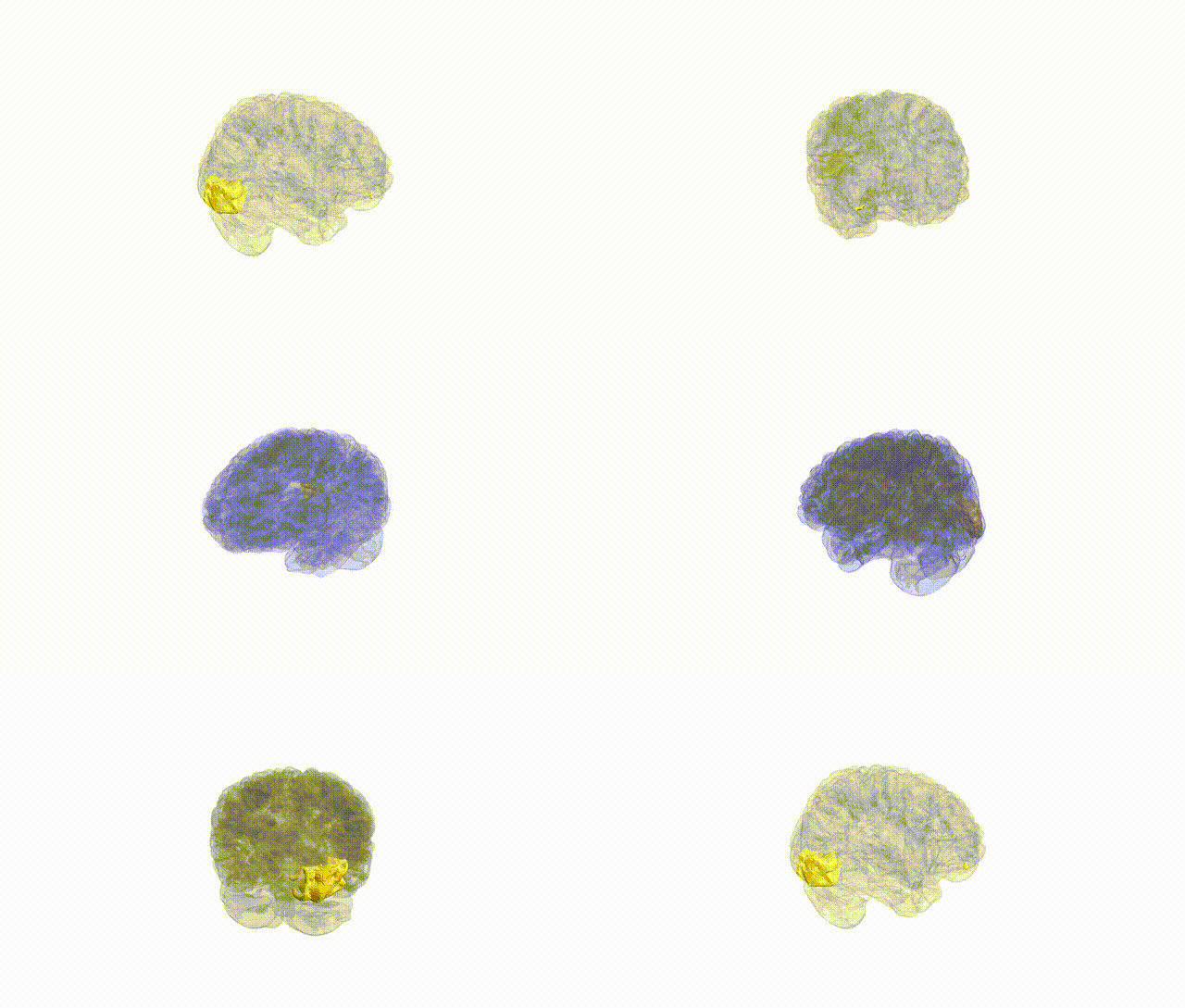
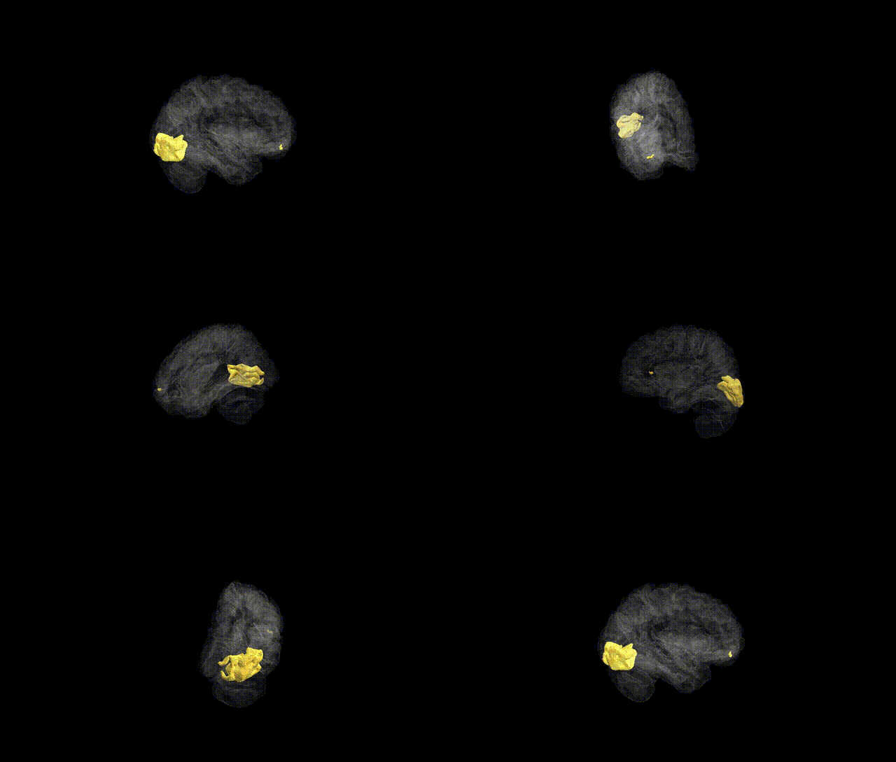
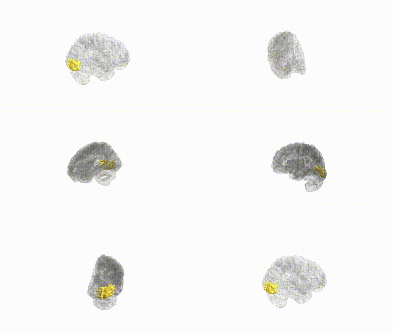
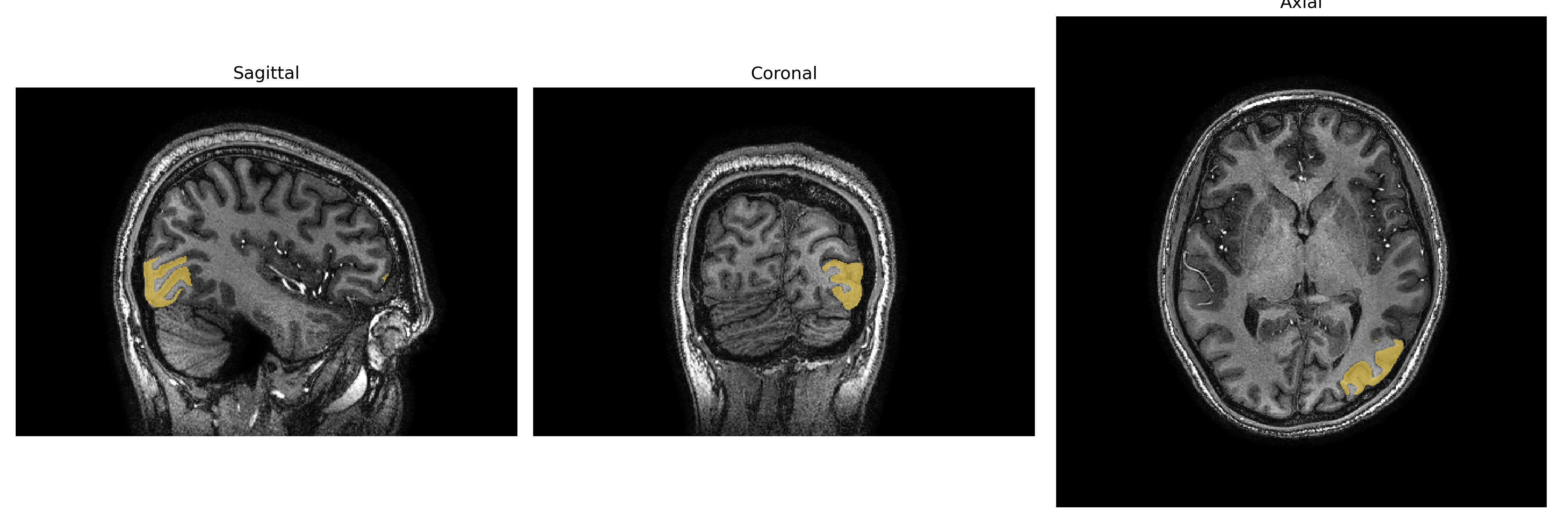
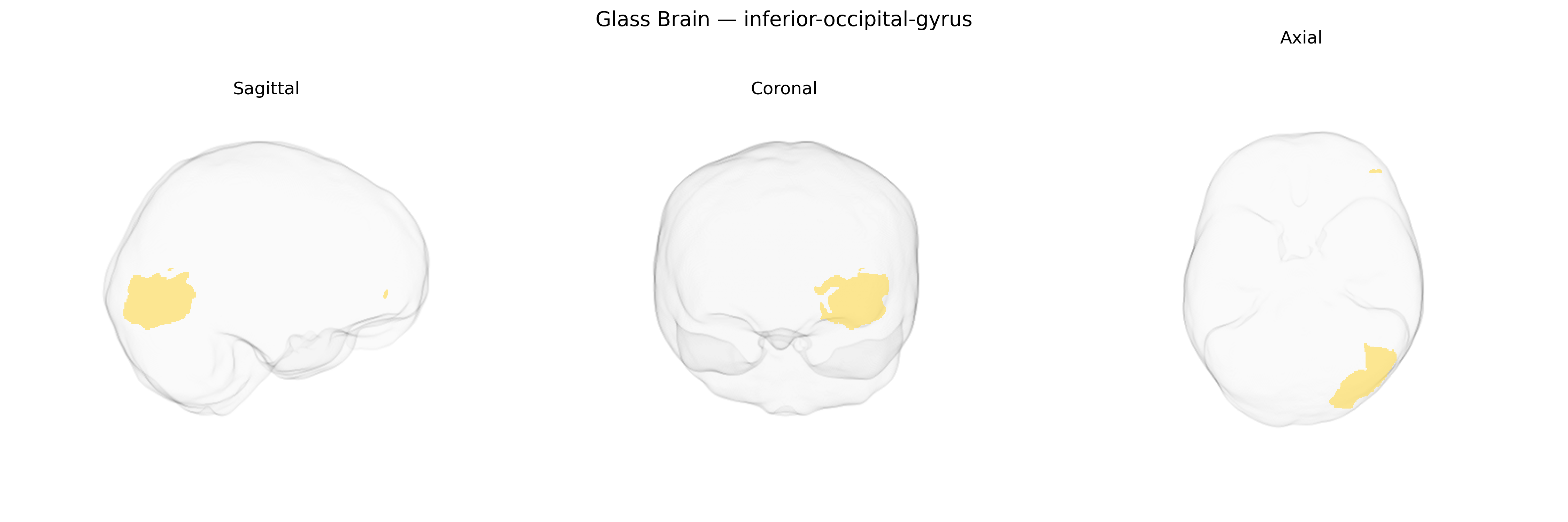
The Left inferior-occipital-gyrus is a cortical region on the ventral surface of the occipital lobe, forming part of the visual association cortex involved in early and intermediate stages of visual processing, particularly analysis of shape, edges, and object features. Cytoarchitectonically, it lies adjacent to and partially overlaps with extrastriate visual areas, contributing to pathways that project anteriorly toward the temporal lobe (ventral “what” stream) for object recognition and category-specific processing. This gyrus receives input from primary and secondary visual areas and participates in integrating low-level visual information into more complex representations used in recognition of objects, faces, and written words. There is no direct link; see the related Occipital lobe.
Genetic associations specifically targeting the Left inferior-occipital-gyrus (L IOG) as defined in the brainCOLOR atlas are sparse, but several large-scale imaging genetics and GWAS studies of cortical thickness, surface area, and volume implicate this region and adjacent occipital cortex in visual processing, face perception, and neurodevelopmental liability. Variants near or within genes involved in synaptic development and axon guidance (e.g., NRXN1, CNTNAP2, DCC, and genes in the semaphorin and ephrin pathways) have shown associations with occipital lobe morphology, though not always isolated to the L IOG. Occipital measures that include the inferior occipital gyrus have been linked in GWAS to general cognitive ability, educational attainment, and psychiatric traits such as schizophrenia and autism spectrum disorder, largely through polygenic influences on cortical surface area and thickness rather than single strong locus effects. Some studies of face-selective and word-selective occipital regions (overlapping the L IOG and nearby fusiform/occipito-temporal cortex) report genetic correlations with reading ability, social cognition, and risk for developmental dyslexia and autism, but these typically treat the occipital gyrus as part of broader visual networks rather than as an isolated parcel. Overall, current evidence suggests that the L IOG inherits a highly polygenic architecture shared with neighboring visual and temporal regions, contributing modest but distributed genetic effects to visual recognition, higher-order cognition, and vulnerability to neurodevelopmental and psychiatric disorders, with no single disorder or variant uniquely and robustly assigned to this exact atlas-defined region.
Overview generated by GPT-4o (2026).
Region ID: 49
Hemisphere: Left
Atlas: brainCOLOR
Full Quality Version: Download MP4
Full Quality Version: Download MP4
Full Quality Version: Download MP4
Full Quality Version: Download MP4
Wali Sidiqyar*, Gaurav Rudravaram*, Elyssa M. McMaster, Trent M. Schwartz, Adam M. Saunders, Kurt G. Schilling, Bennett A. Landman "Introducing SPINS: A Shared Public Visualization Library of Neuroanatomical Structures." Medical Imaging with Deep Learning- short paper
This resource is licensed under CC0 1.0 Universal (Public Domain).