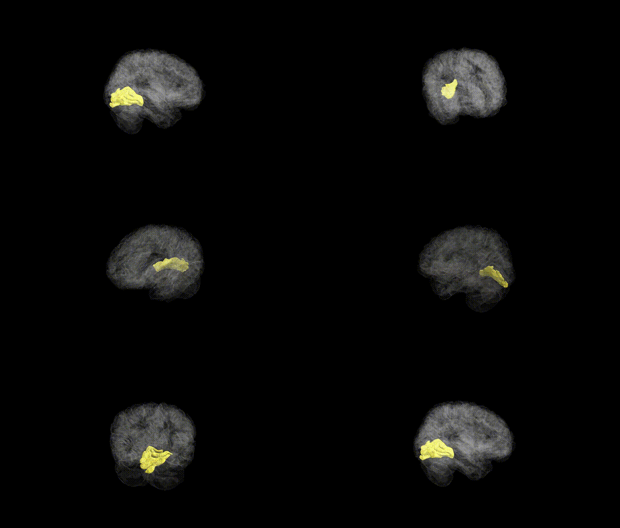
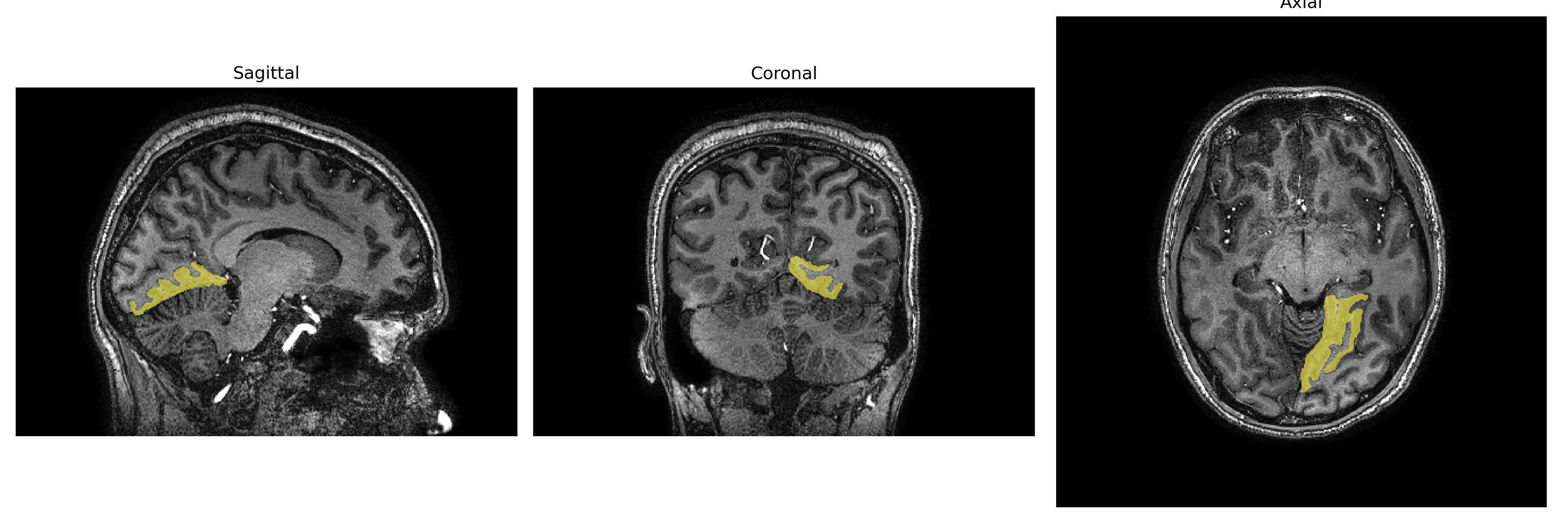
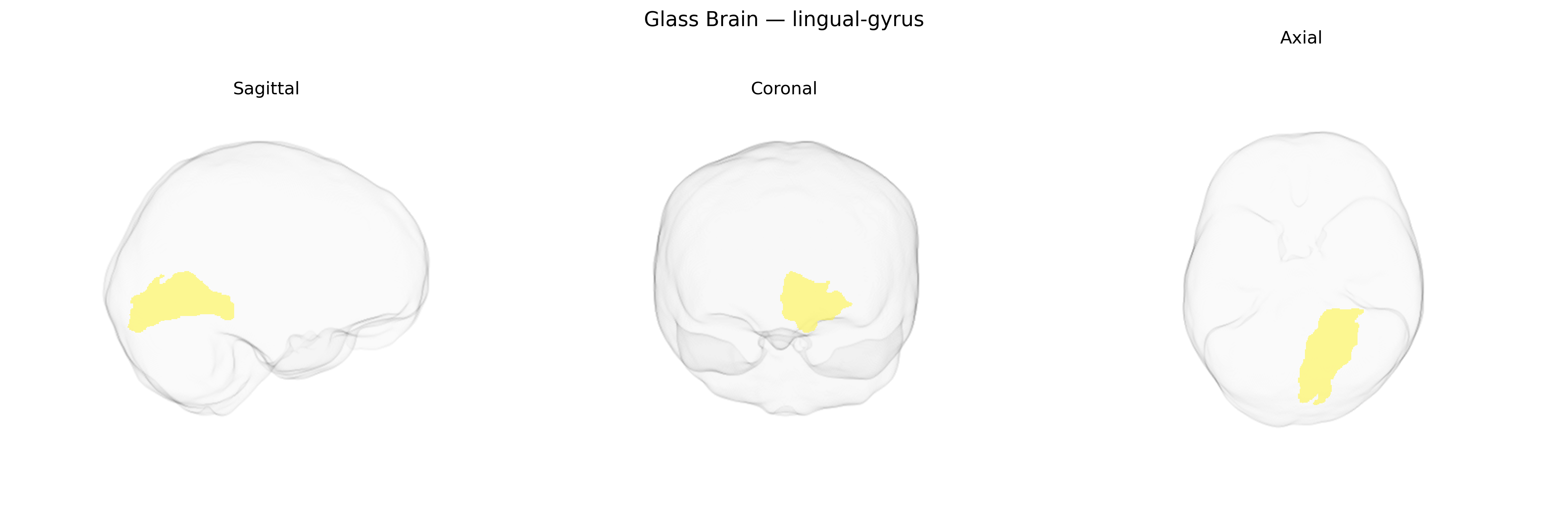
The left lingual gyrus is an occipital lobe structure situated on the medial aspect of the cerebral hemisphere, extending from the calcarine sulcus to the posterior portion of the temporal lobe, and is primarily supplied by branches of the posterior cerebral artery. Cytoarchitectonically, it includes portions of visual association cortex involved in complex processing of visual stimuli, notably color, form, and letter/word recognition, and contributes critically to visual word form processing and reading, particularly in the dominant (usually left) hemisphere. It maintains reciprocal connections with primary visual cortex (V1), higher-order visual association areas, and language-related regions in the temporal and parietal lobes, allowing integration of visual information with linguistic and semantic networks. Functional imaging frequently implicates the left lingual gyrus in visual imagery, orthographic processing, and certain aspects of episodic memory retrieval that rely on visual cues.
Lingual gyrus
Genetic associations involving the left lingual gyrus, as defined in the brainCOLOR atlas, largely emerge from imaging-genetics and neuropsychiatric GWAS that implicate this occipital–medial temporal region in visual processing, reading, and affective function. Twin and SNP-heritability studies show that lingual gyrus volume and cortical thickness are moderately to highly heritable, with common variants in genes involved in neurodevelopment, synaptic plasticity, and myelination (for example, pathways including BDNF signaling, cell-adhesion molecules, and glutamatergic transmission) contributing to structural variation, though robust single-locus hits specific to the left lingual gyrus are sparse. Large-scale brain-structure GWAS (e.g., ENIGMA, UK Biobank) report loci that influence occipital and ventral visual-stream regions, including lingual gyrus metrics, some of which overlap with genetic risk factors for schizophrenia, major depressive disorder, bipolar disorder, and autism spectrum disorder, consistent with case–control MRI findings of altered lingual morphology or activation in these conditions. Additional associations link lingual gyrus activation or structure to polygenic risk scores for depression, anxiety, and psychosis, and to traits such as cognitive performance, educational attainment, reading and language skills, and susceptibility to visual hallucinations and migraine aura; however, these findings generally reflect shared polygenic influences on distributed networks rather than effects uniquely confined to the left lingual gyrus.
Overview generated by GPT-4o (2026).
Region ID: 53
Hemisphere: Left
Atlas: brainCOLOR
Full Quality Version: Download MP4
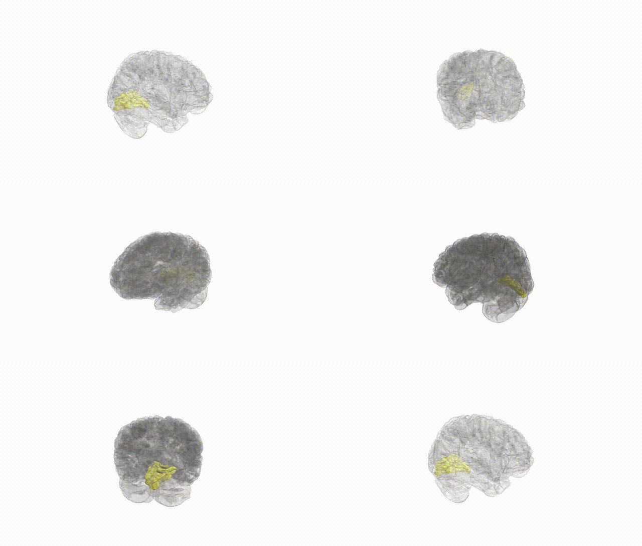
Full Quality Version: Download MP4
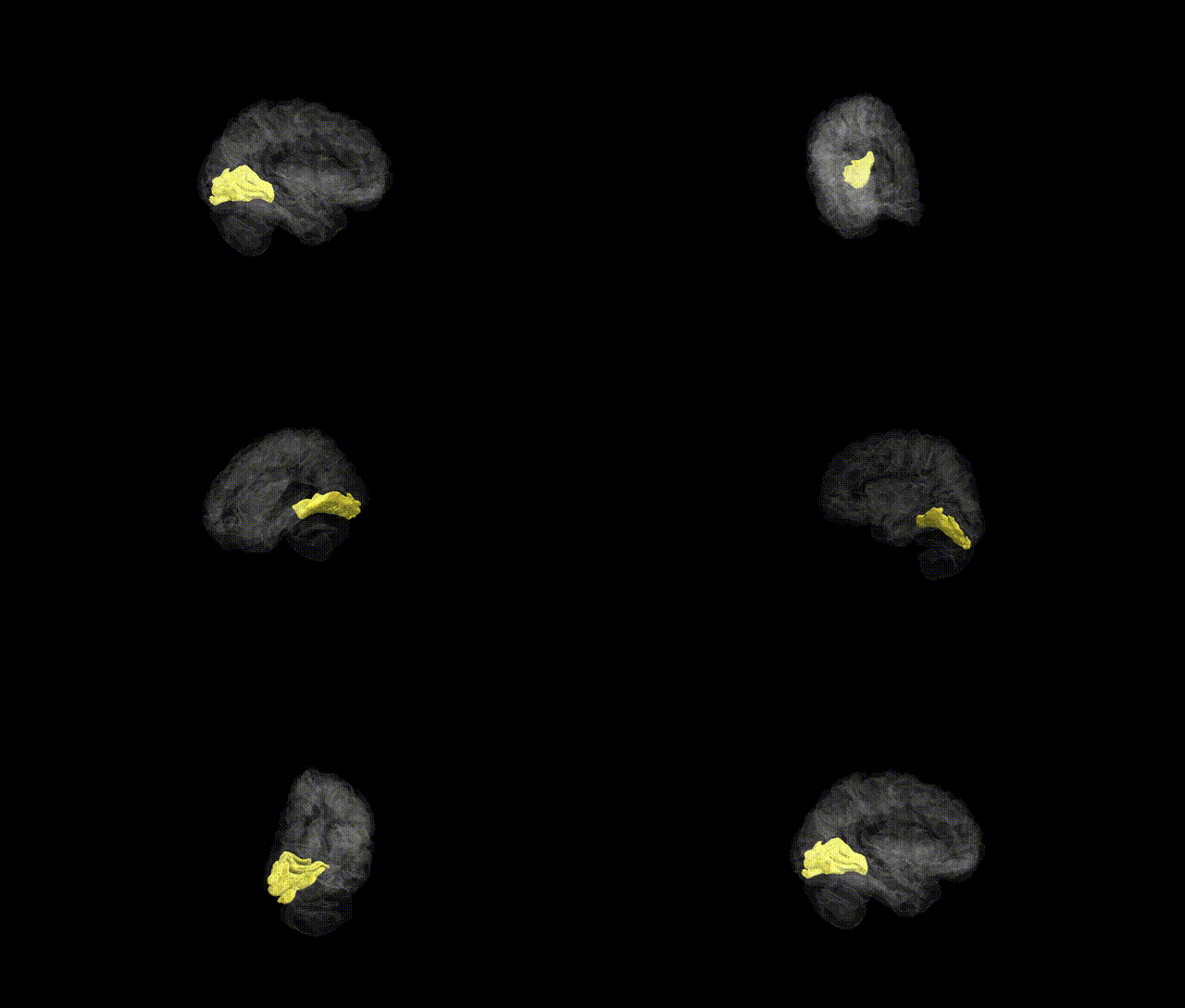
Full Quality Version: Download MP4
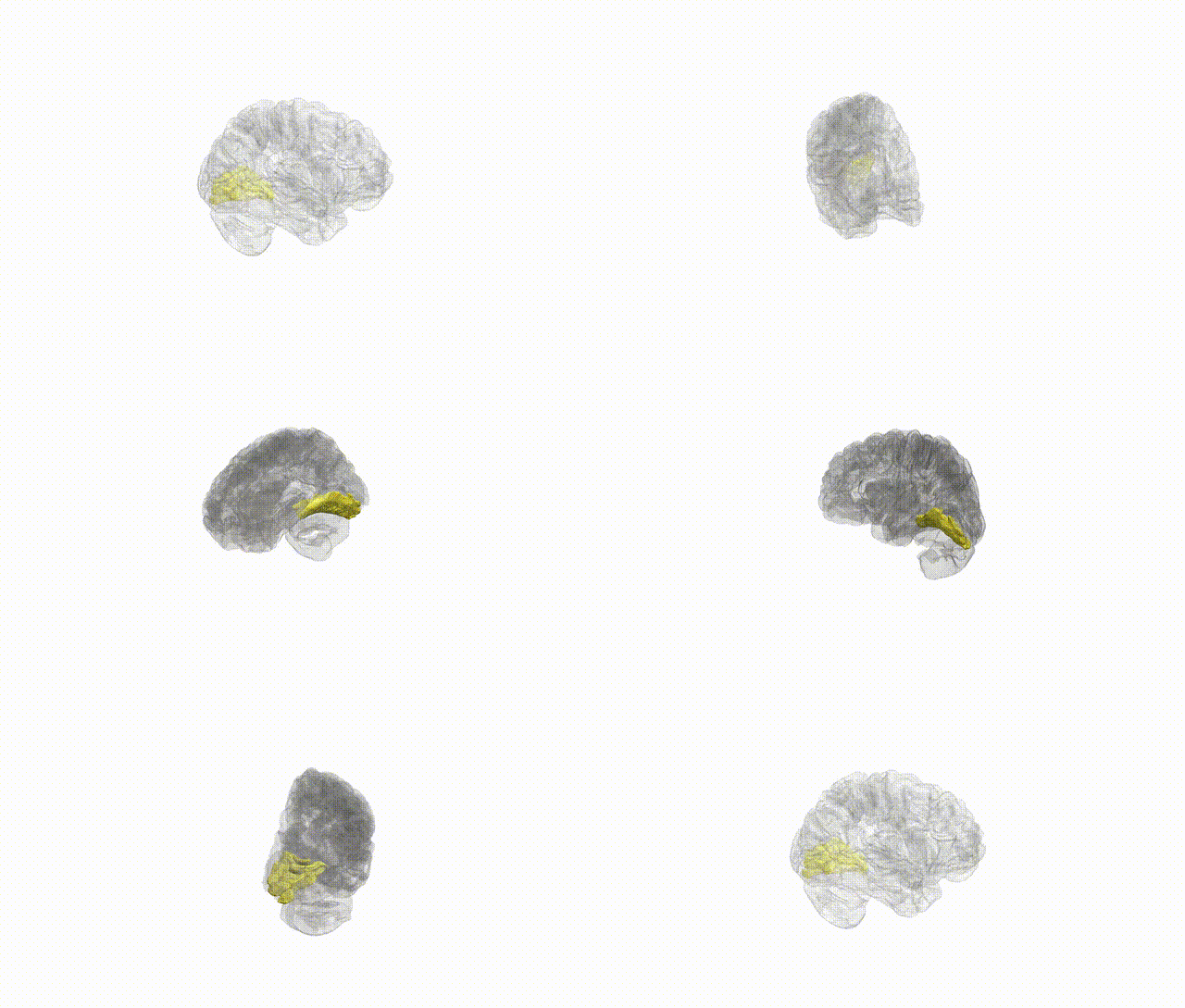
Full Quality Version: Download MP4
Wali Sidiqyar*, Gaurav Rudravaram*, Elyssa M. McMaster, Trent M. Schwartz, Adam M. Saunders, Kurt G. Schilling, Bennett A. Landman "Introducing SPINS: A Shared Public Visualization Library of Neuroanatomical Structures." Medical Imaging with Deep Learning- short paper
This resource is licensed under CC0 1.0 Universal (Public Domain).