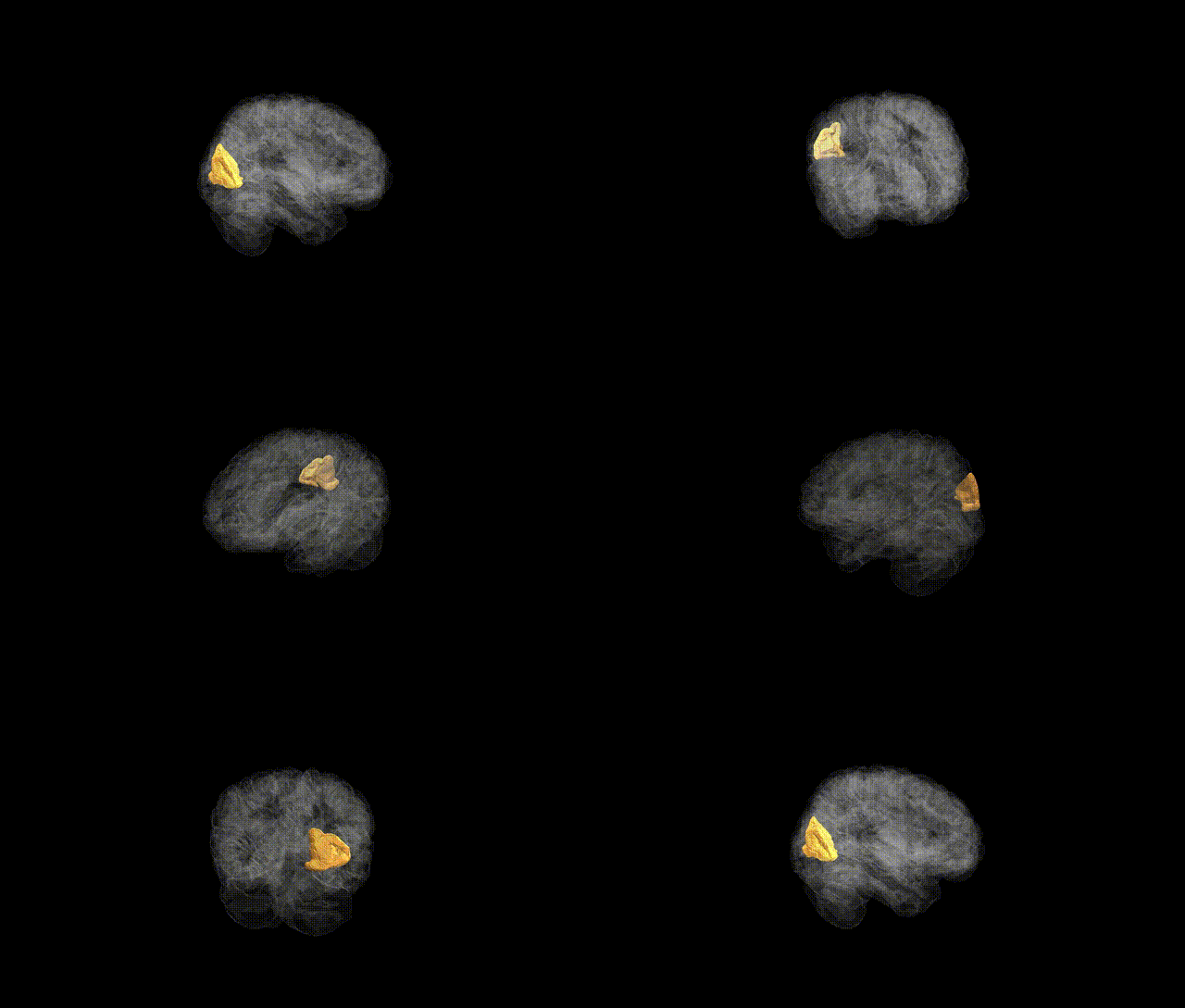
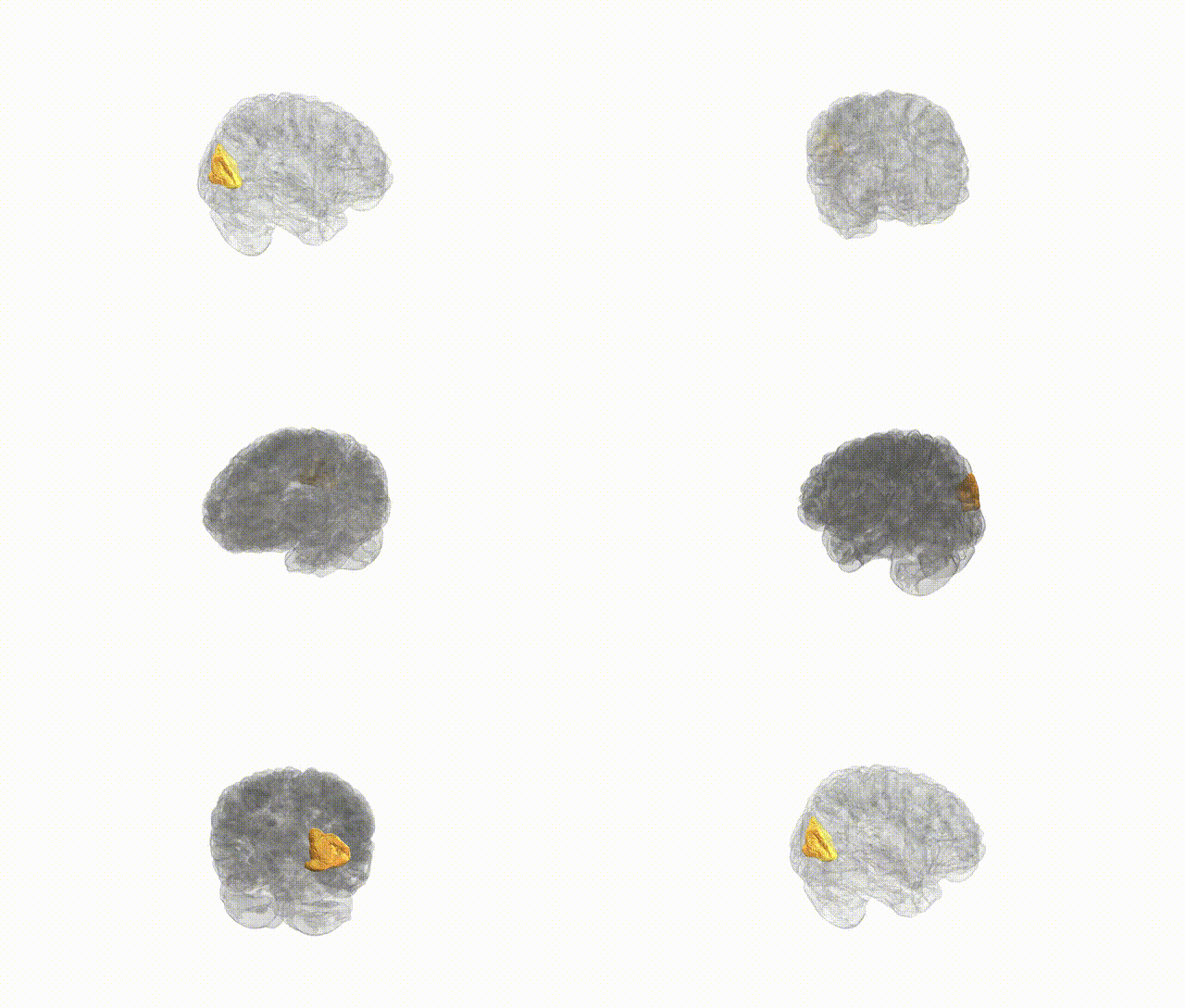
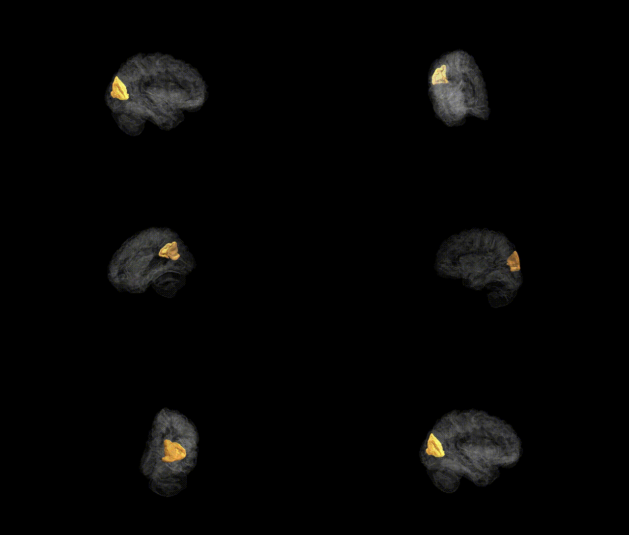
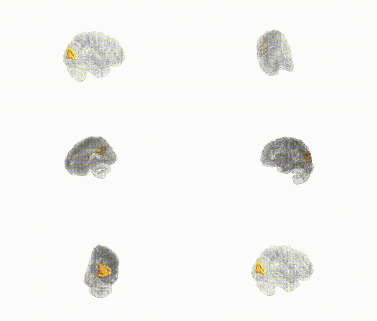
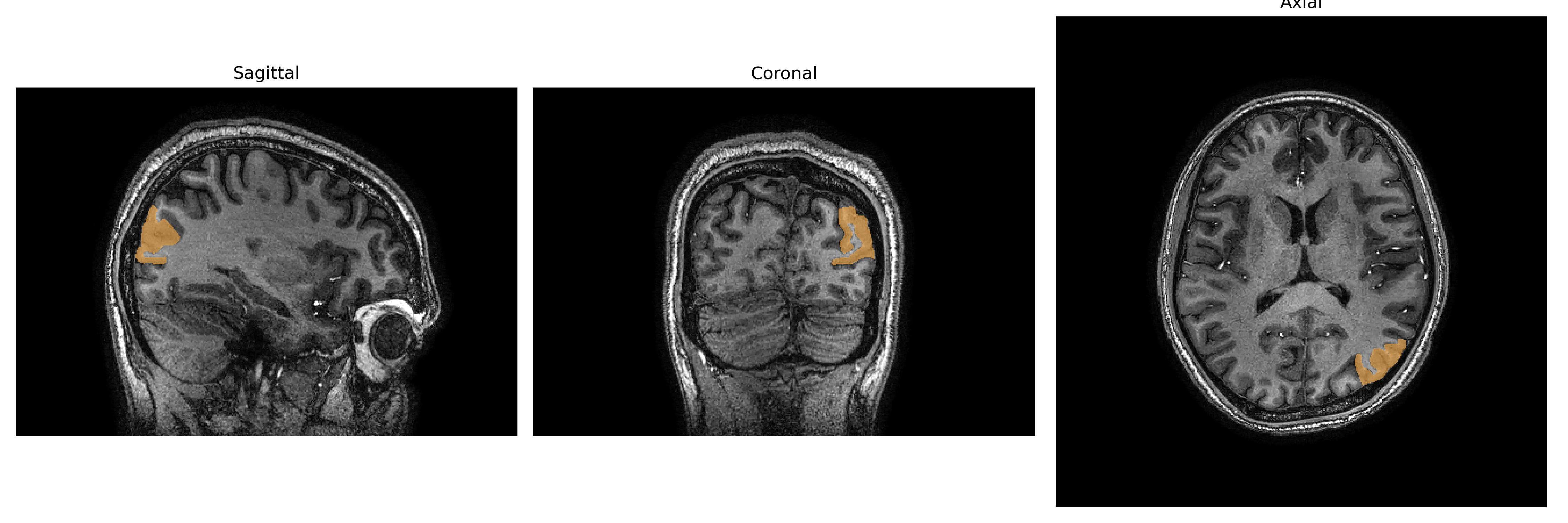
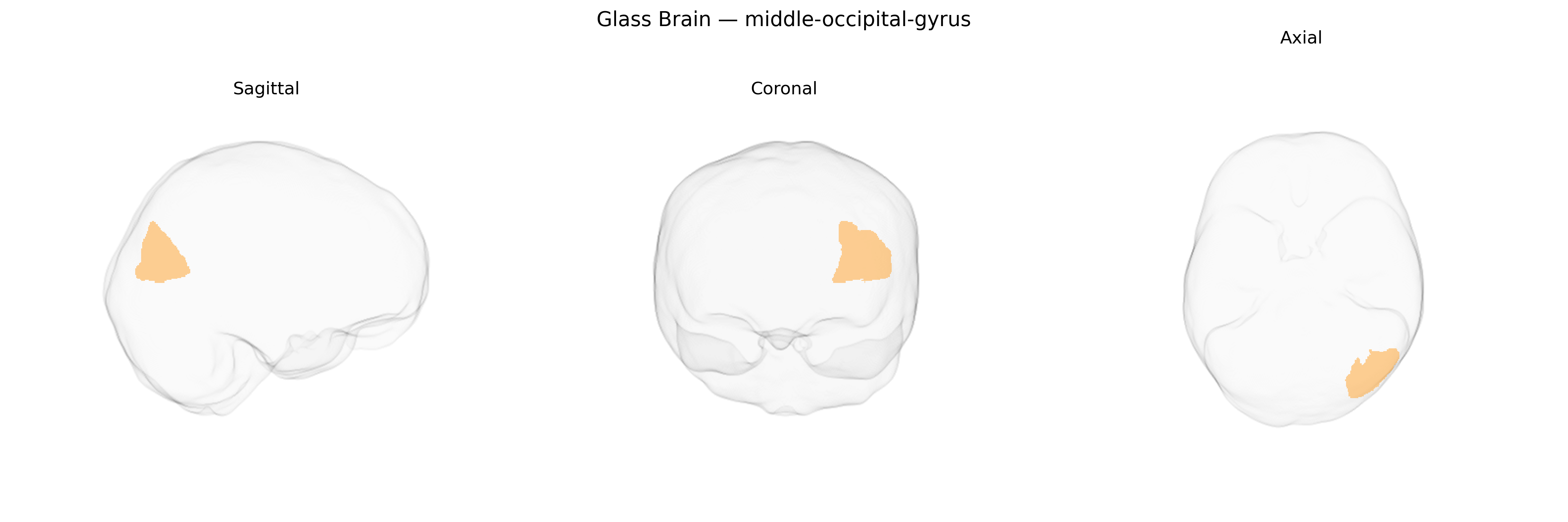
The Left middle-occipital gyrus is a lateral occipital cortical region located on the convexity of the occipital lobe, typically bounded superiorly by the superior occipital gyrus and inferiorly by the inferior occipital gyrus, and extending anteriorly toward the occipito-temporal junction. It is composed predominantly of visual association cortex (Brodmann areas primarily in the 18/19 range), integrating visual input from primary visual areas and contributing to higher-order processing of form, motion, and spatial aspects of the visual field, mainly representing the right visual hemifield. This region participates in dorsal and ventral visual stream interactions, supporting visuospatial analysis, object recognition, and visually guided behavior, and is structurally connected with parietal and temporal association cortices as well as subcortical visual pathways. There is no direct link for “middle occipital gyrus,” but it is part of the Occipital lobe.
The left middle occipital gyrus, a core visual-association region in the brainCOLOR Atlas, has been implicated in multiple imaging-genetics and GWAS studies that link its structure and function to common genetic variation affecting visual processing, higher-order cognition, and neuropsychiatric risk. Large-scale brain MRI GWAS consortia (e.g., ENIGMA, UK Biobank–based studies) have identified SNPs near genes involved in neurodevelopment, synaptic function, and axon guidance—such as variants in or near regulatory regions of MAPT, HMGA2, and several chromosome 14 and 17 loci—that show associations with occipital cortical thickness, surface area, or volume, including the middle occipital gyrus. Polygenic risk scores for schizophrenia, bipolar disorder, and major depressive disorder have been associated with subtle structural differences in occipital regions, suggesting that some psychiatric risk architectures converge on visual-association cortex, though effects are typically small and distributed. Additionally, genetic risk for educational attainment and general cognitive ability has been linked to occipital morphology and activation patterns in visual and reading tasks, implicating the left middle occipital gyrus in reading and visuospatial functions influenced by common variants. Occipital cortex, including the middle occipital gyrus, is also implicated in migraine and visual aura susceptibility, with GWAS-identified variants in genes related to vascular tone and cortical excitability (e.g., near TRPM8, LRP1) contributing to altered visual-cortical responsiveness. Overall, available evidence supports a polygenic architecture in which numerous small-effect variants, many shared across cognitive and psychiatric traits, shape the structure and function of the left middle occipital gyrus rather than any single strong locus uniquely defining this region.
Overview generated by GPT-4o (2026).
Region ID: 63
Hemisphere: Left
Atlas: brainCOLOR
Full Quality Version: Download MP4
Full Quality Version: Download MP4
Full Quality Version: Download MP4
Full Quality Version: Download MP4
Wali Sidiqyar*, Gaurav Rudravaram*, Elyssa M. McMaster, Trent M. Schwartz, Adam M. Saunders, Kurt G. Schilling, Bennett A. Landman "Introducing SPINS: A Shared Public Visualization Library of Neuroanatomical Structures." Medical Imaging with Deep Learning- short paper
This resource is licensed under CC0 1.0 Universal (Public Domain).