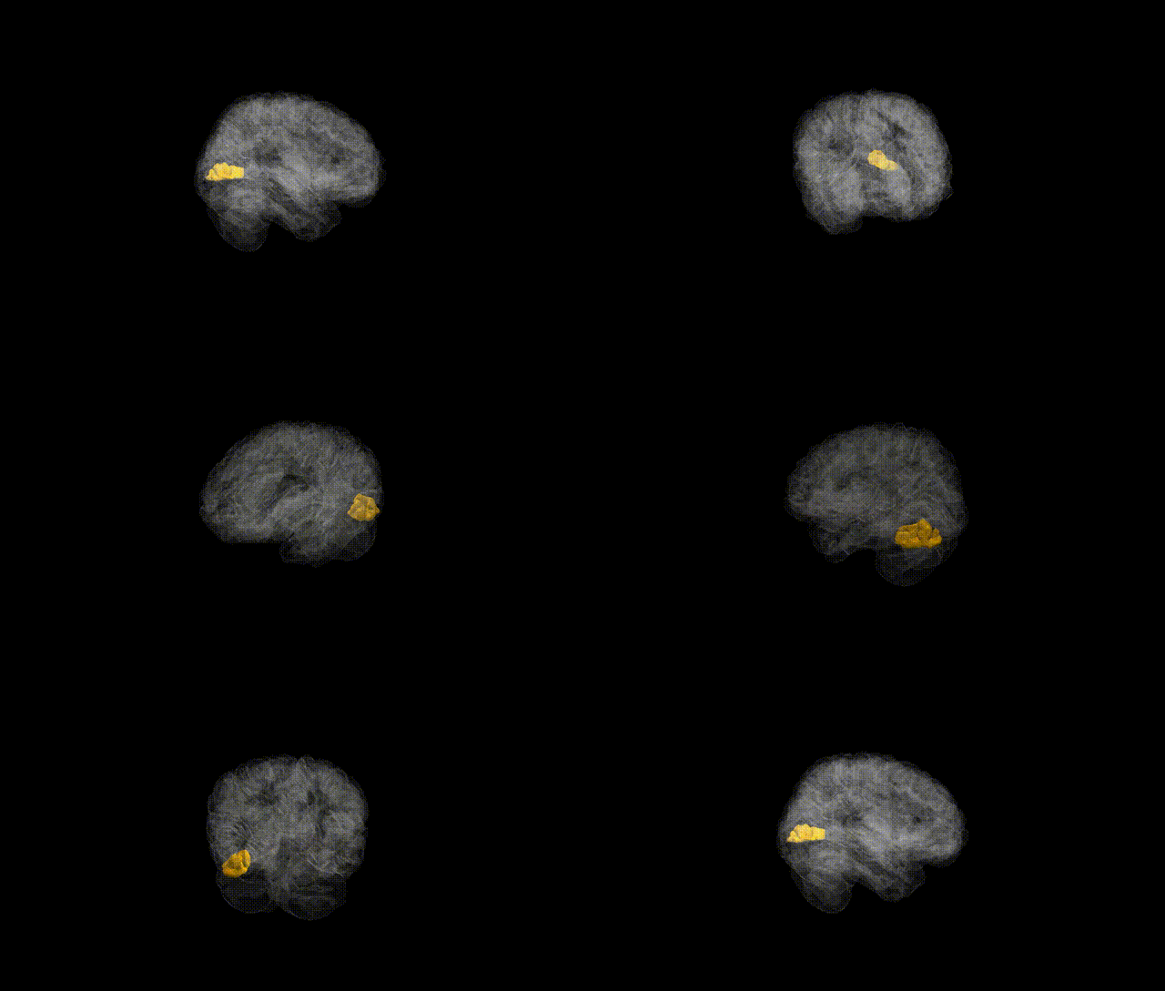
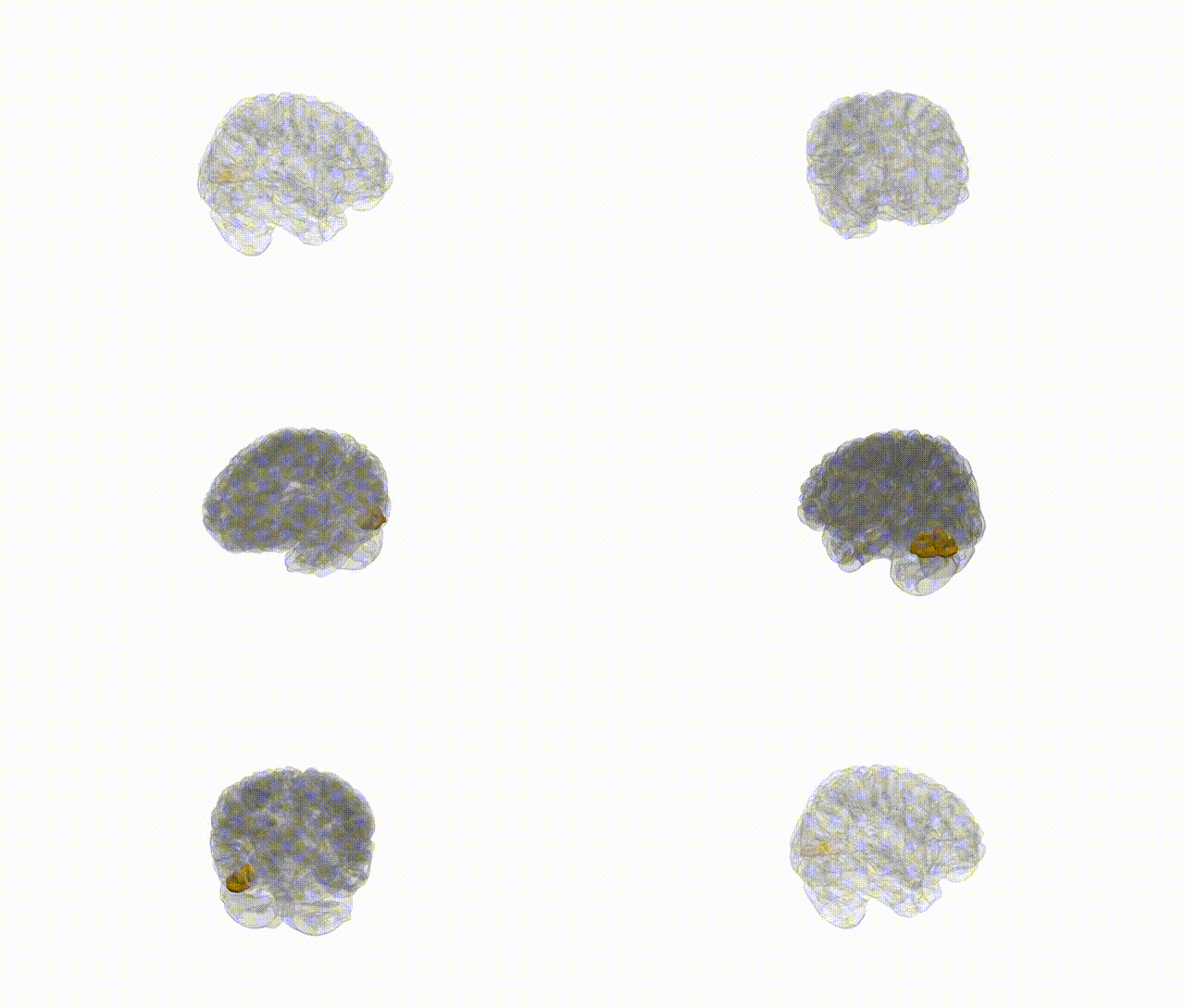
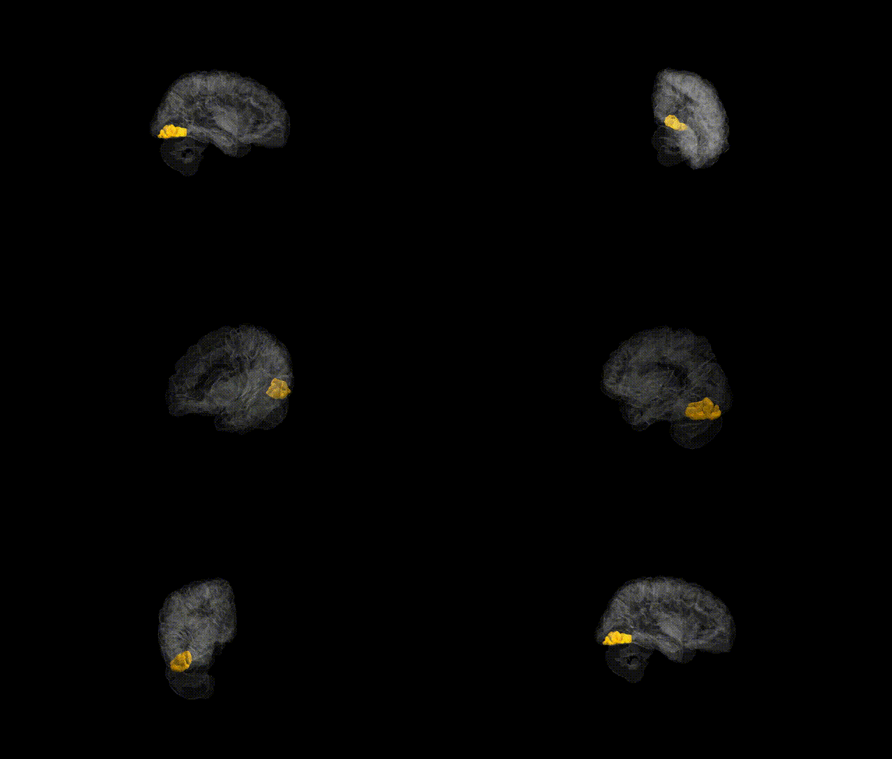
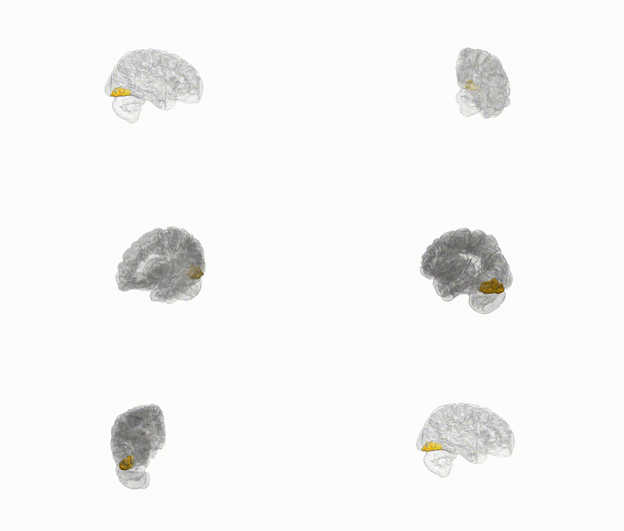
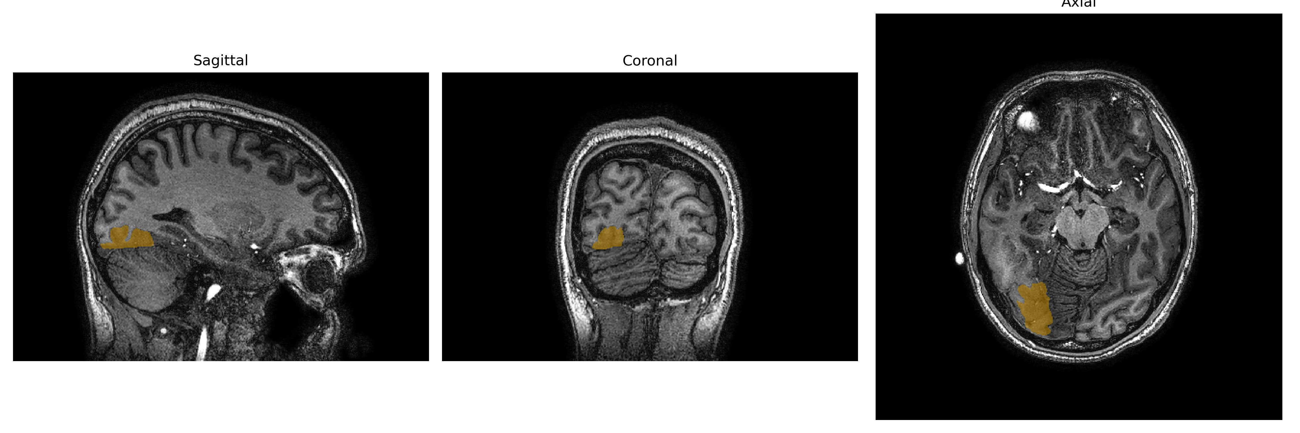
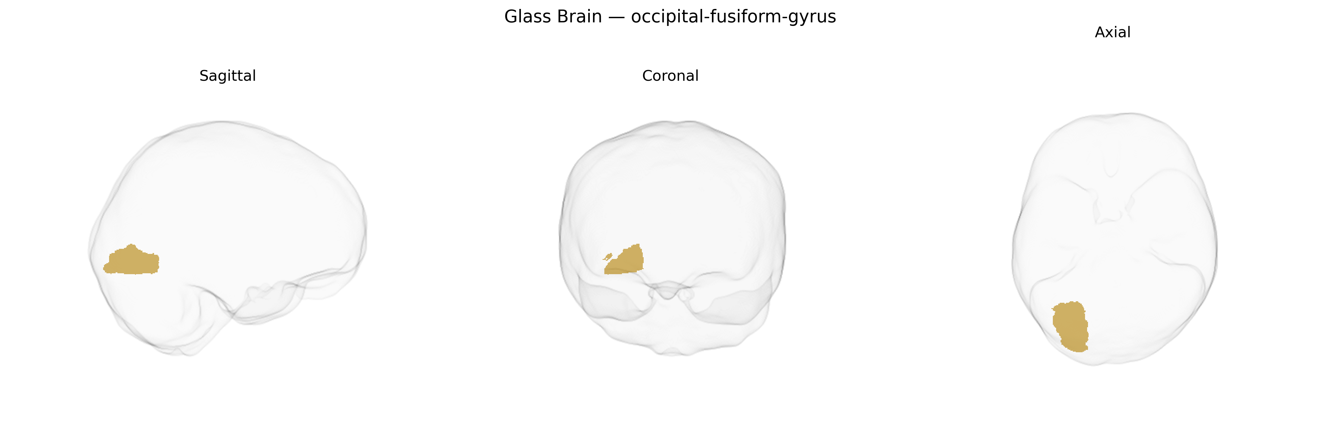
The Right occipital-fusiform gyrus, as defined in the brainCOLOR atlas, corresponds to the posterior portion of the fusiform gyrus extending into the occipital lobe, forming part of the ventral visual stream. This region participates in higher-order visual processing, including the analysis of complex object features such as shape, texture, and fine-grained visual details, and contributes to category-selective responses (e.g., faces, words, or other visually meaningful stimuli) depending on its precise subregion and connectivity. It receives input from early visual cortices and projects to more anterior temporal regions, integrating visual information with semantic and memory-related processes. Anatomically, the occipital-fusiform area lies on the basal surface of the temporal and occipital lobes, bordered medially by the collateral sulcus and laterally by the occipitotemporal sulcus, and is often considered part of the broader fusiform gyrus complex. A closely related structure with a detailed description is the Fusiform gyrus.
Genetic associations with the right occipital-fusiform gyrus, as defined in the brainCOLOR Atlas, largely emerge from imaging genetics and GWAS of cortical structure and function rather than from region-specific candidate-gene studies. Large-scale neuroimaging GWAS (e.g., ENIGMA and UK Biobank) have identified common variants in genes involved in neurodevelopment and synaptic function—such as KIAA0586, DAAM1, and MIR138-2—associated with occipito-temporal cortical thickness, surface area, and gyrification, which encompass fusiform and adjacent occipital regions. Polygenic risk scores for neurodevelopmental and psychiatric disorders, including autism spectrum disorder, schizophrenia, and major depressive disorder, have been correlated with structural and functional variation in occipito-temporal regions that overlap the right occipital-fusiform gyrus, consistent with this region’s role in visual processing and social perception. GWAS of face recognition ability and reading-related traits (including dyslexia risk) have repeatedly implicated voxels in the right fusiform and occipitotemporal cortex, although these studies typically report functional clusters rather than atlas-specific parcels, so the mapping to the brainCOLOR right occipital-fusiform region is approximate. Overall, current evidence links genetic variation influencing cortical development, synaptic signaling, and neuropsychiatric liability to structural and functional variability in occipito-fusiform territories, but no robust, uniquely specific gene set has been established for the right occipital-fusiform gyrus as delineated by the brainCOLOR Atlas.
Overview generated by GPT-4o (2026).
Region ID: 76
Hemisphere: Right
Atlas: brainCOLOR
Full Quality Version: Download MP4
Full Quality Version: Download MP4
Full Quality Version: Download MP4
Full Quality Version: Download MP4
Wali Sidiqyar*, Gaurav Rudravaram*, Elyssa M. McMaster, Trent M. Schwartz, Adam M. Saunders, Kurt G. Schilling, Bennett A. Landman "Introducing SPINS: A Shared Public Visualization Library of Neuroanatomical Structures." Medical Imaging with Deep Learning- short paper
This resource is licensed under CC0 1.0 Universal (Public Domain).