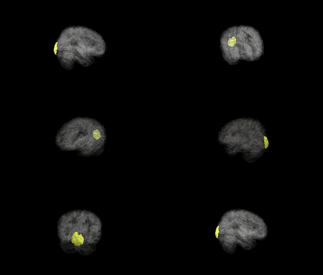
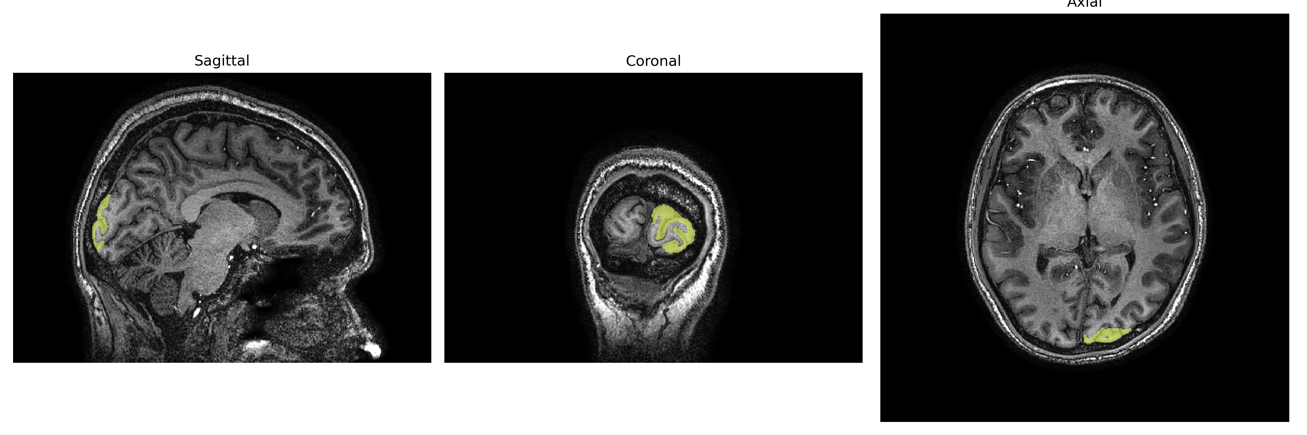
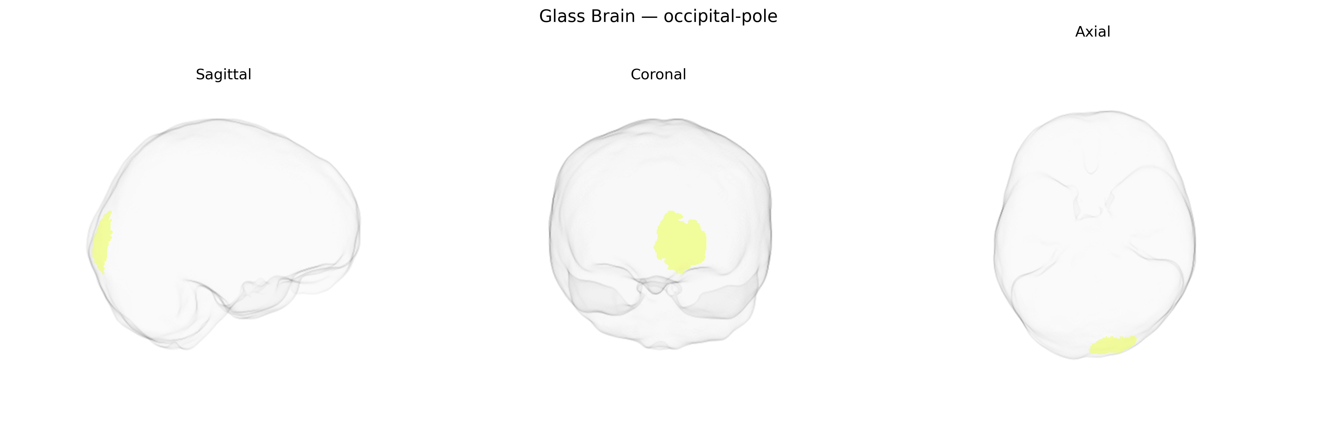
The left occipital pole is the most posterior portion of the left occipital lobe, forming part of the primary and early visual cortical areas that receive and process visual input from the contralateral (right) visual field via projections from the lateral geniculate nucleus of the thalamus. It contains regions of the striate (V1) and peristriate cortex involved in initial encoding of basic visual features such as orientation, contrast, spatial frequency, and simple motion, which are then relayed to higher-order dorsal and ventral visual streams for complex processing, including object recognition and spatial analysis. Lesions restricted to the left occipital pole typically produce right-sided central or paracentral visual field defects and can contribute to visual perceptual disorders depending on the extent of damage and involvement of adjoining visual association areas. There is no direct link for “left occipital pole,” but it is part of the Occipital lobe.
The left occipital pole, encompassing primary and early visual cortices in the brainCOLOR atlas, has been implicated in several large-scale imaging genetics and GWAS efforts that link variation in occipital surface area, cortical thickness, and volume to common genetic variants, particularly in genes involved in neurodevelopment, synaptic function, and axon guidance. ENIGMA and UK Biobank–based studies of cortical morphology have identified loci influencing occipital pole metrics in or near genes such as MCPH1 and HMGA2 (general cortical size and surface area), and more broadly within pathways regulating neuronal proliferation and cortical arealization, though region-specific loci for the left occipital pole are typically modest and often shared with other visual or posterior cortical regions. Occipital pole structure and activation patterns have also been genetically correlated with traits such as general cognitive ability, educational attainment, and visual acuity, as well as with psychiatric and neurodevelopmental conditions including schizophrenia, autism spectrum disorder, and major depressive disorder, where polygenic risk scores and GWAS hits for these disorders show enriched associations with visual and association cortex measures rather than uniquely with the left occipital pole. In addition, migraine—especially migraine with aura—and certain forms of epilepsy (e.g., occipital lobe epilepsy) show converging evidence of altered occipital structure and excitability, and GWAS findings for migraine implicate genes affecting cortical excitability and vascular regulation that may influence occipital pole physiology. Overall, genetic associations with the left occipital pole are consistent with its role as part of a broader visual and multimodal network, with most known variants exerting small, distributed effects rather than conferring a specific, isolated genetic signature on this single brainCOLOR-defined region.
Overview generated by GPT-4o (2026).
Region ID: 75
Hemisphere: Left
Atlas: brainCOLOR
Full Quality Version: Download MP4
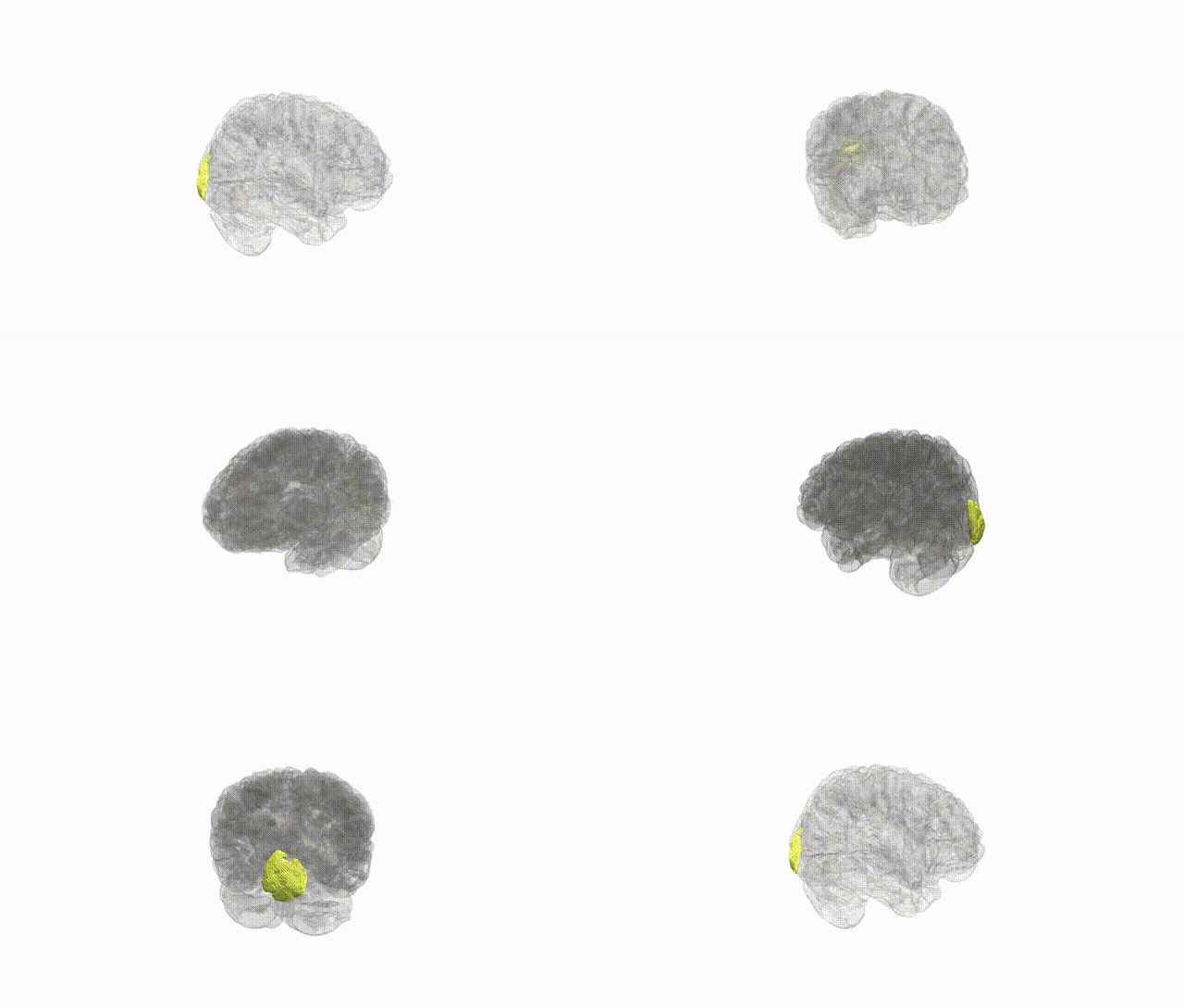
Full Quality Version: Download MP4
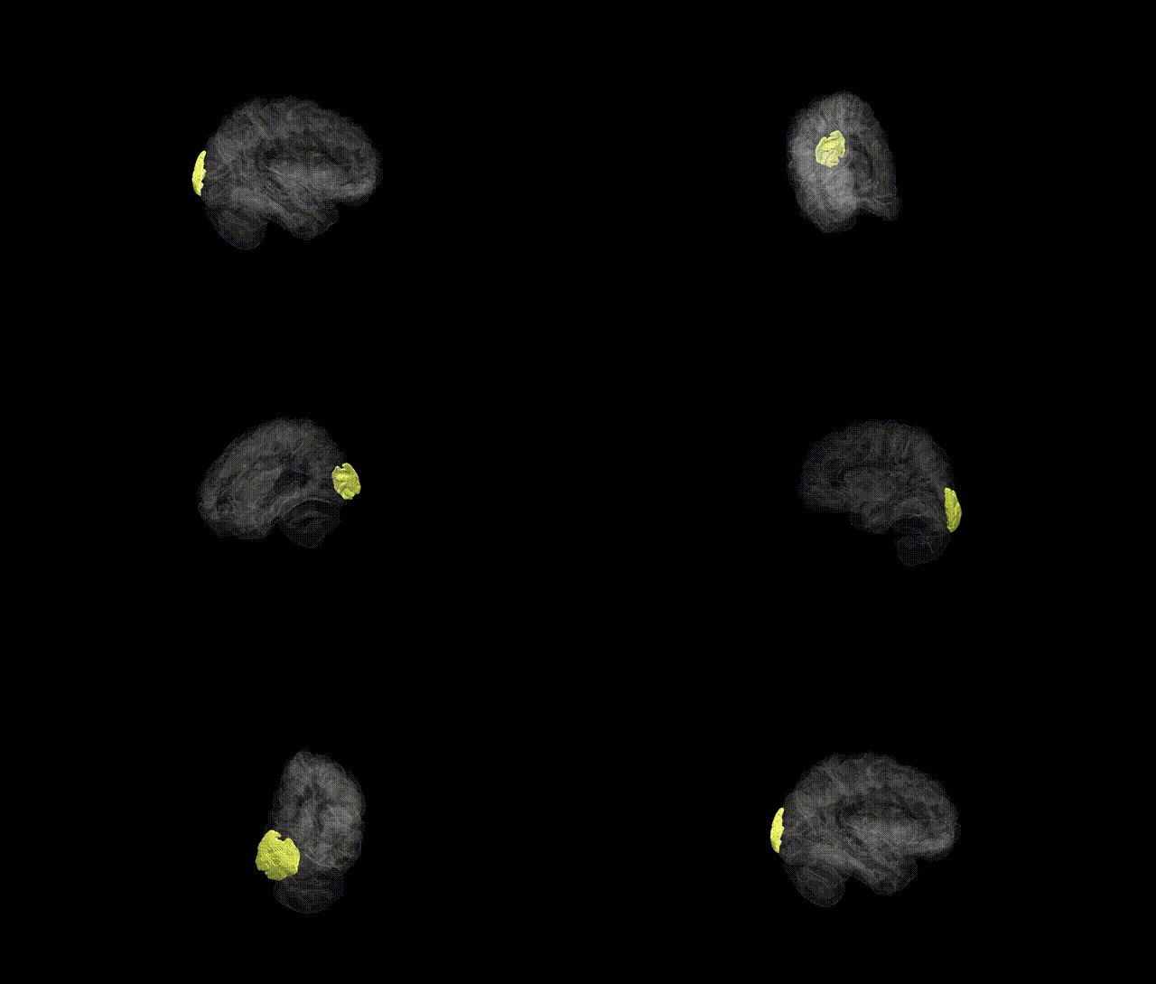
Full Quality Version: Download MP4
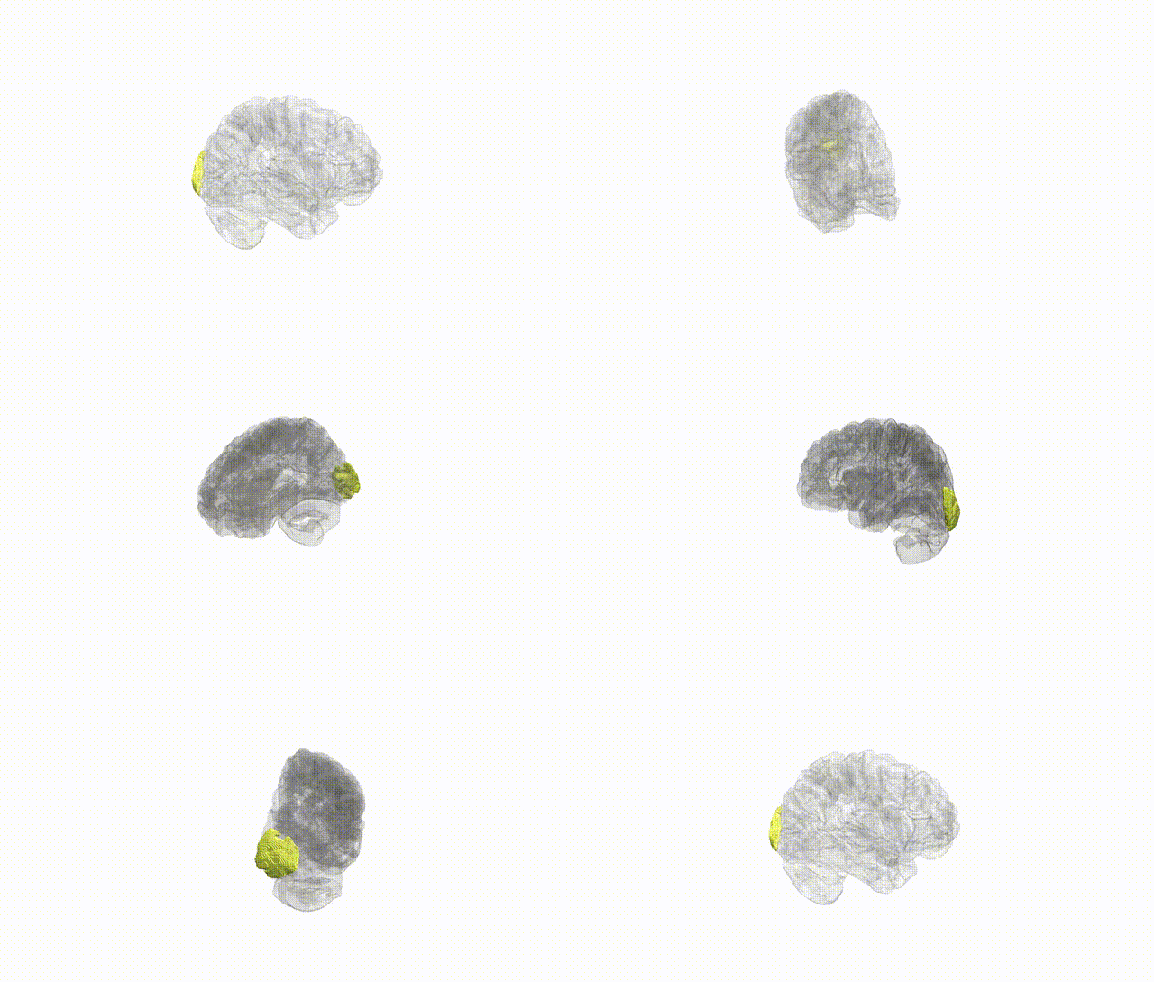
Full Quality Version: Download MP4
Wali Sidiqyar*, Gaurav Rudravaram*, Elyssa M. McMaster, Trent M. Schwartz, Adam M. Saunders, Kurt G. Schilling, Bennett A. Landman "Introducing SPINS: A Shared Public Visualization Library of Neuroanatomical Structures." Medical Imaging with Deep Learning- short paper
This resource is licensed under CC0 1.0 Universal (Public Domain).