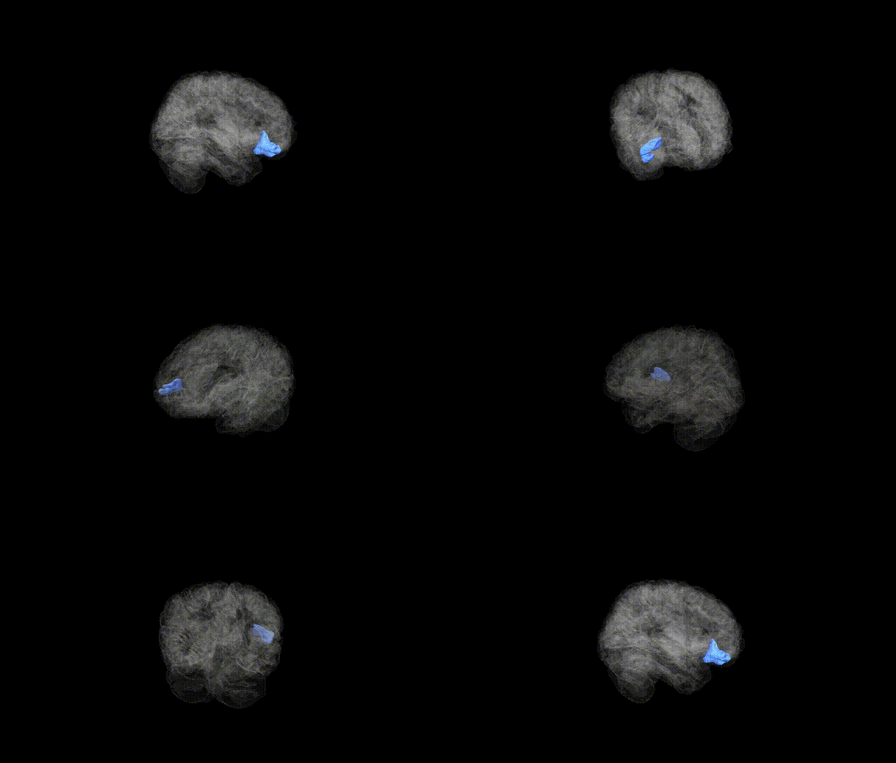
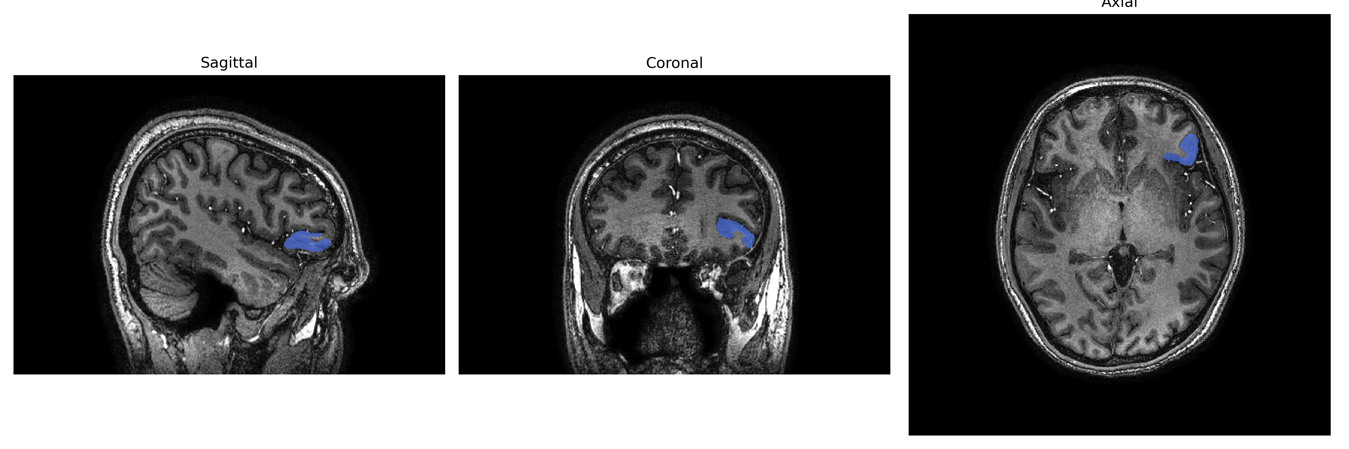
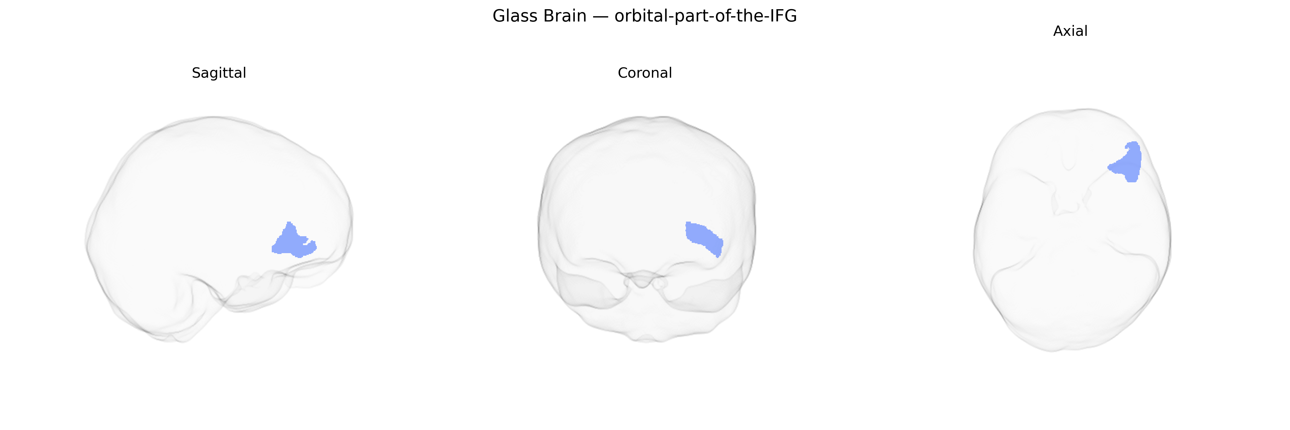
The Left orbital part of the inferior frontal gyrus (IFG) is a ventral subdivision of the inferior frontal cortex located on the orbital surface of the frontal lobe, anterior and inferior to the insula and bordering the lateral orbitofrontal cortex. It lies inferior to the pars triangularis and pars opercularis of the IFG and is positioned above the orbital plate of the frontal bone, overlying the anterior cranial fossa. Cytoarchitectonically, it corresponds largely to portions of Brodmann areas 11 and 47, and it is strongly interconnected with limbic, temporal, and orbitofrontal regions. Functionally, this region is implicated in the integration of emotional and reward-related information with higher-order cognition, including aspects of decision making, valuation, and social-emotional processing, and may contribute to language-related functions via its connections with other IFG subdivisions. There is no direct Wikipedia article specific to the “orbital part of the IFG”; a closely related and encompassing region is the Inferior frontal gyrus.
The left orbital part of the inferior frontal gyrus (IFG), as defined in parcellations such as the brainCOLOR Atlas, has emerged in genetic and imaging–genetics studies as part of a broader prefrontal network implicated in language, social cognition, and affective regulation, with several genome-wide association studies (GWAS) linking its morphology or function to common variants. Large-scale ENIGMA and UK Biobank analyses of cortical thickness, surface area, and volume have identified heritable variation in inferior frontal and orbitofrontal regions associated with loci near genes involved in neurodevelopment and synaptic function (for example, variants in or near MIR137, DLG2, and genes in glutamatergic and GABAergic pathways), although effects are typically distributed and not specific to a single subregion. Functionally, the broader left IFG (including orbital parts) has been implicated in GWAS of reading and language-related traits, with risk variants for dyslexia and related learning disorders mapping onto networks that prominently include left IFG; polygenic scores for educational attainment and cognitive performance also correlate with structural measures in prefrontal regions encompassing the orbital IFG. Psychiatric GWAS for major depressive disorder, bipolar disorder, schizophrenia, ADHD, and autism spectrum disorder consistently highlight genes whose expression or regulatory architecture is enriched in frontal cortex, and case–control imaging–genetics work frequently shows altered left orbitofrontal/IFG volume or activity associated with disorder-linked polygenic risk scores, particularly for depression and schizophrenia. In addiction and impulsivity-related traits, variants in dopaminergic and serotonergic genes and polygenic liability for substance use disorders have been associated with altered activation or connectivity of orbitofrontal–IFG circuitry during reward and inhibition tasks. Overall, available evidence indicates that the left orbital IFG is a heritable, genetically influenced node within fronto-limbic and language networks, with genetic associations emerging primarily through distributed polygenic effects rather than region-specific “single-gene” links.
Overview generated by GPT-4o (2026).
Region ID: 81
Hemisphere: Left
Atlas: brainCOLOR
Full Quality Version: Download MP4
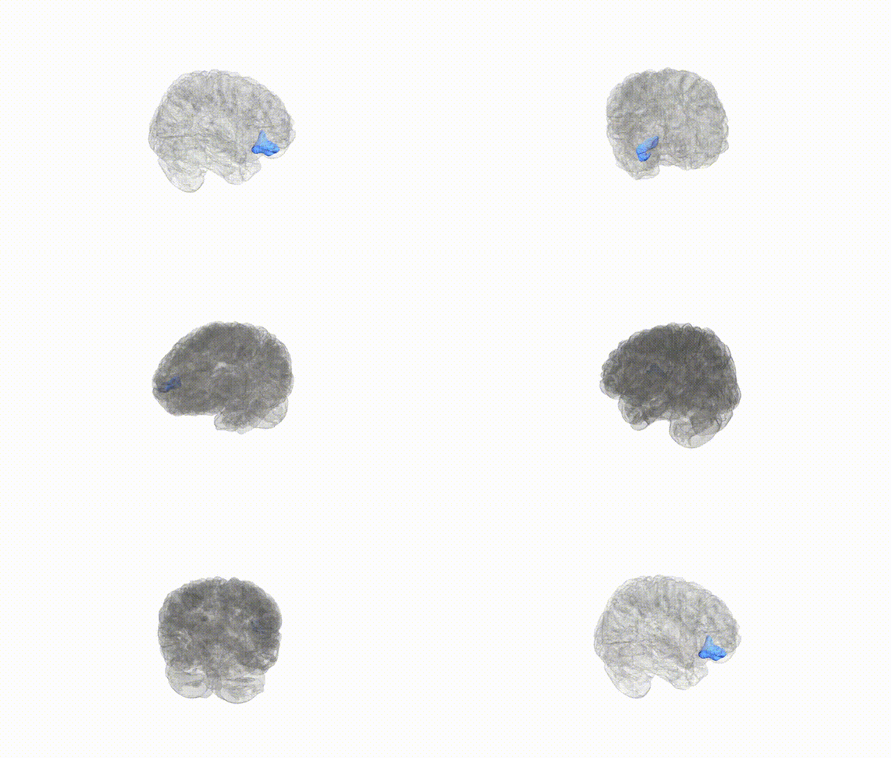
Full Quality Version: Download MP4
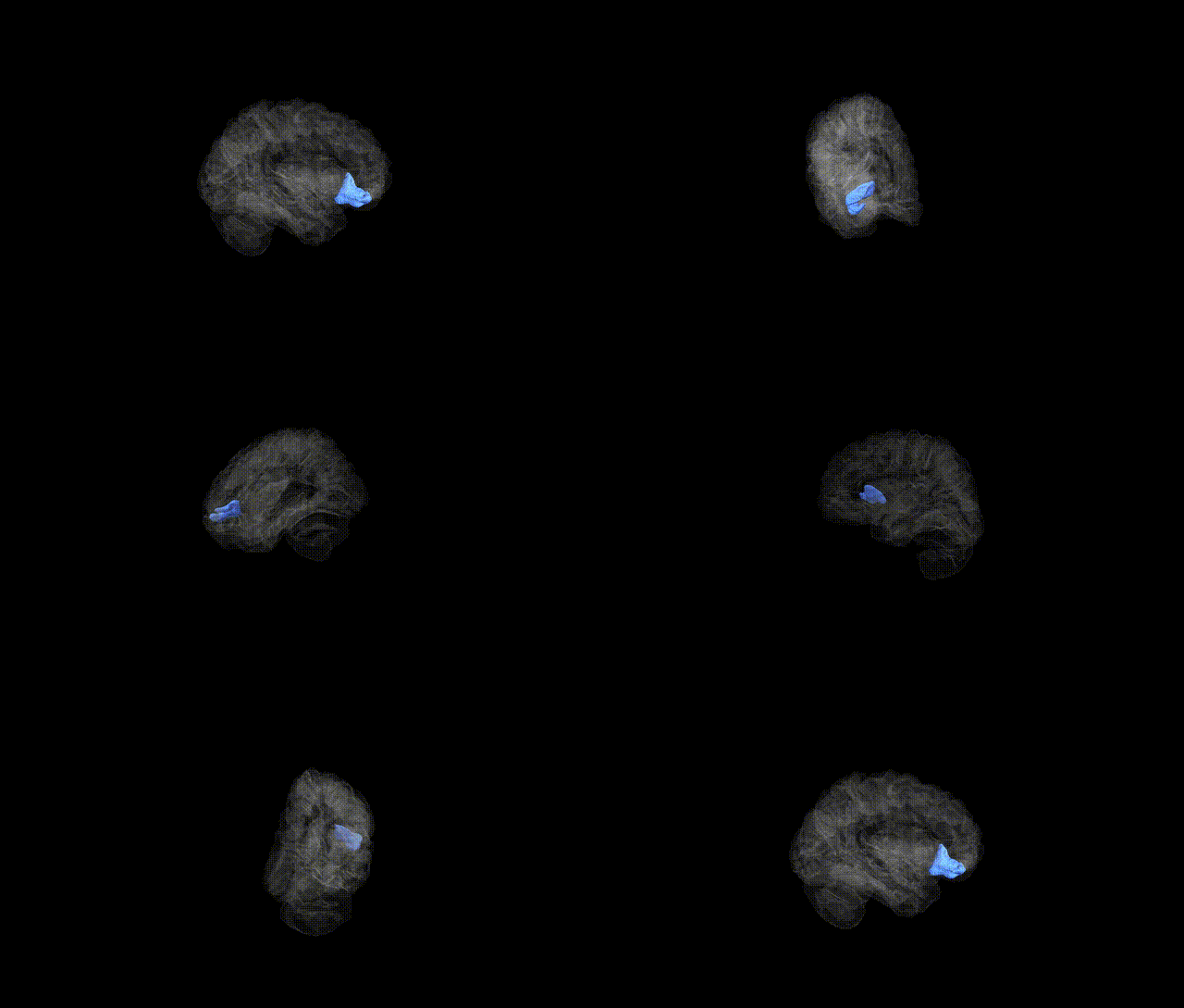
Full Quality Version: Download MP4
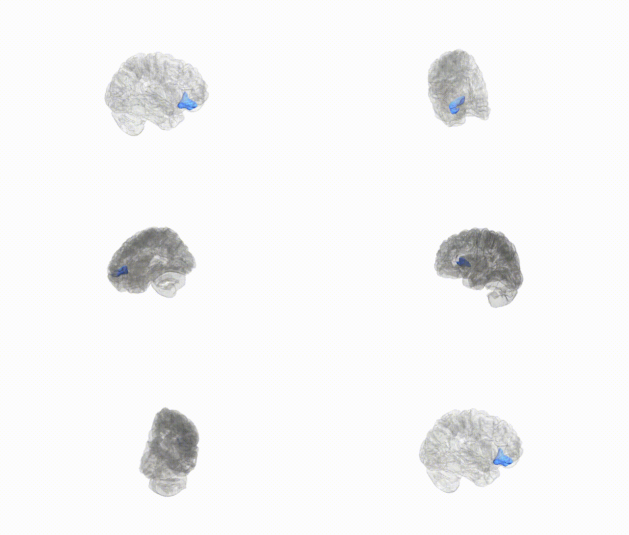
Full Quality Version: Download MP4
Wali Sidiqyar*, Gaurav Rudravaram*, Elyssa M. McMaster, Trent M. Schwartz, Adam M. Saunders, Kurt G. Schilling, Bennett A. Landman "Introducing SPINS: A Shared Public Visualization Library of Neuroanatomical Structures." Medical Imaging with Deep Learning- short paper
This resource is licensed under CC0 1.0 Universal (Public Domain).