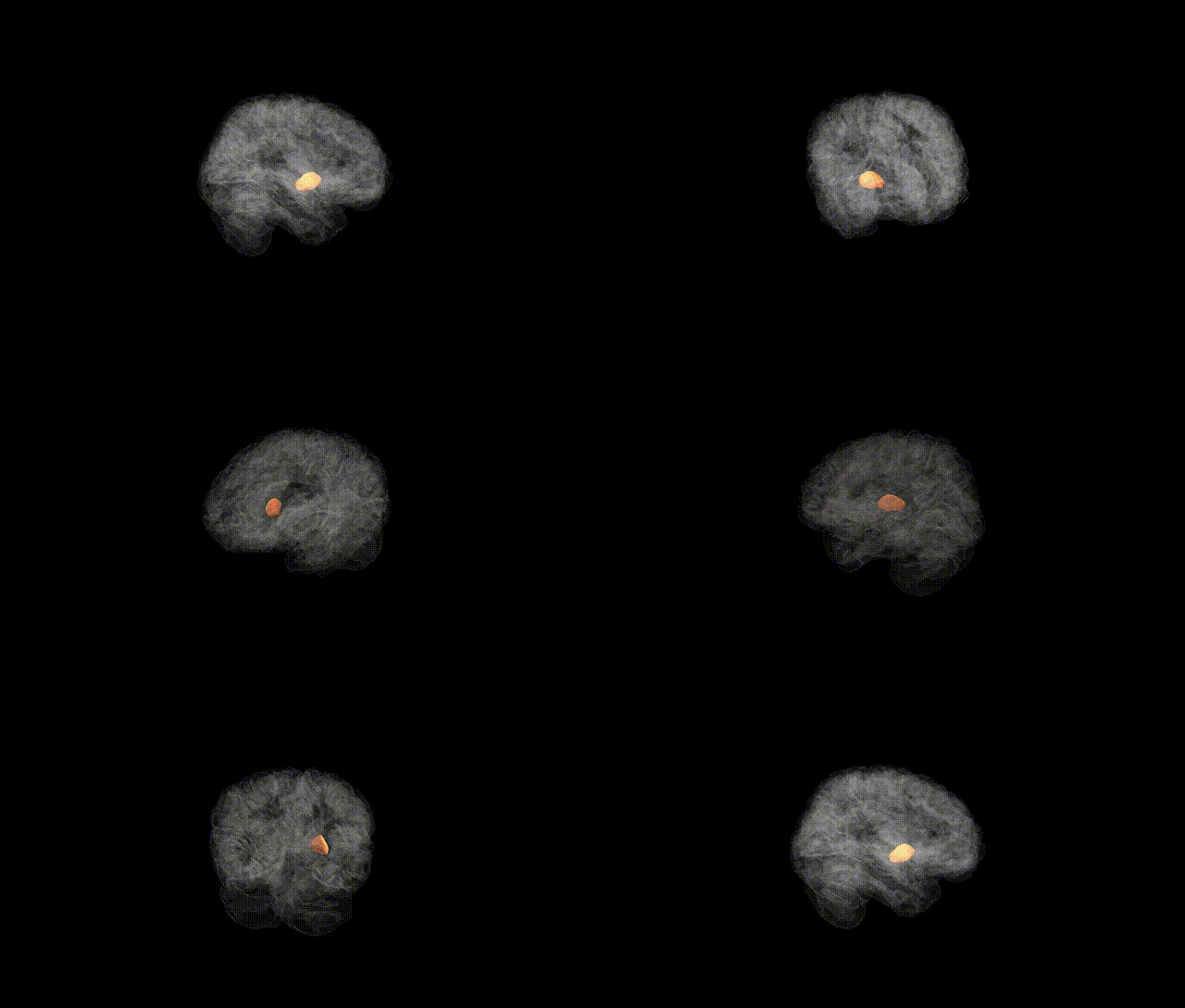
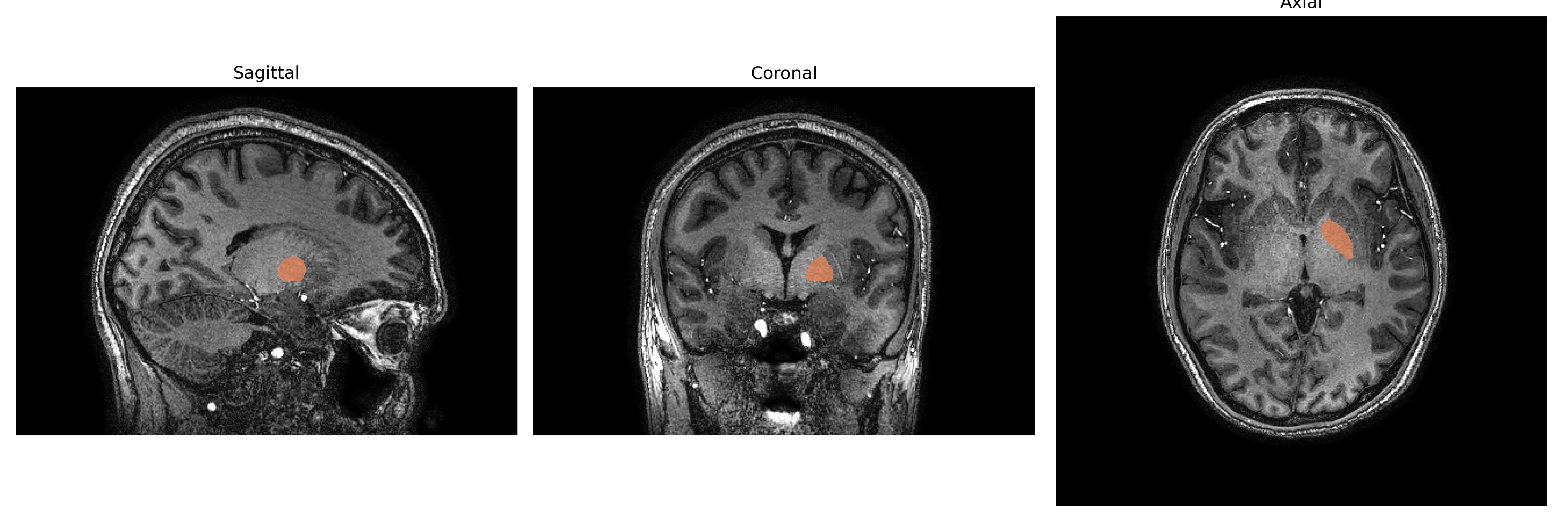
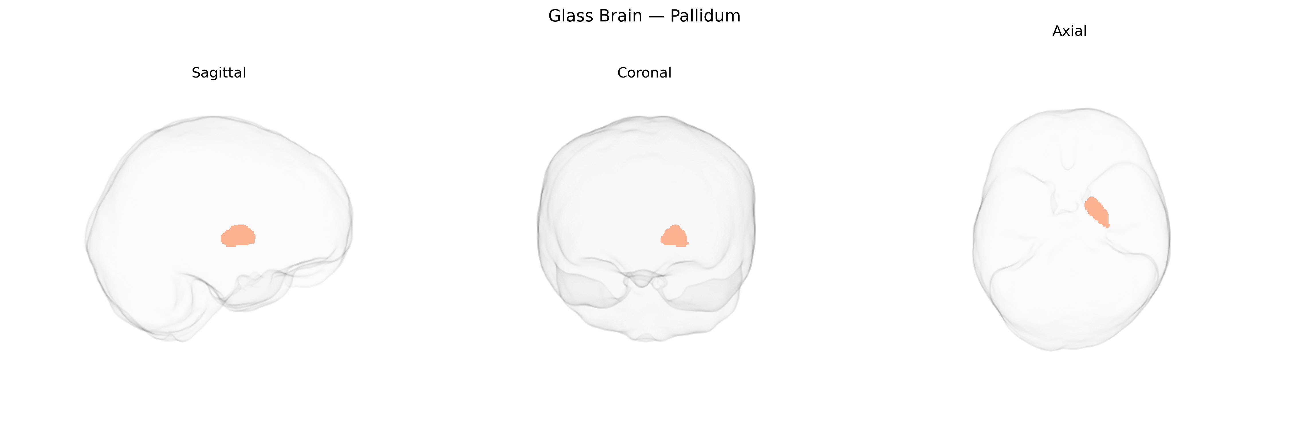
The Left Pallidum is the left-sided component of the globus pallidus, a major basal ganglia nucleus located in the ventral part of the forebrain, medial to the putamen and lateral to the internal capsule. Composed of internal (medial) and external (lateral) segments, it is primarily formed by GABAergic projection neurons that exert inhibitory control over thalamocortical and brainstem motor pathways. Functionally, the Left Pallidum participates in the regulation of voluntary movement, muscle tone, and motor learning as part of cortico–basal ganglia–thalamocortical loops, and also contributes to aspects of motivation and reward processing through its connections with limbic and associative circuits. Pallidal dysfunction is implicated in movement disorders such as Parkinson’s disease, dystonia, and chorea, where alterations in its inhibitory output lead to characteristic motor symptoms. There is no direct Wikipedia article for the “Left Pallidum” as a standalone entry; a related structure and functional context is described under Globus pallidus.
The left pallidum, a basal ganglia structure prominent in motor, cognitive, and reward circuits, shows significant heritability and has been implicated in several genome-wide association studies (GWAS) examining subcortical brain volumes, psychiatric disease, and neurodegeneration. Large imaging-genetics consortia such as ENIGMA and UK Biobank–based studies have identified common variants near or within genes involved in neurodevelopment, synaptic function, and dopaminergic or glutamatergic signaling that influence pallidal volume or morphology, including loci in or near genes like KTN1, DCC, and putative neuronal growth and guidance genes, though effect sizes are generally modest and often shared across multiple subcortical structures rather than specific to the pallidum alone. Pallidal measures have been associated with polygenic risk scores for schizophrenia, major depressive disorder, bipolar disorder, Parkinson’s disease, and Alzheimer’s disease, and several disorder-focused GWAS and imaging–genetics studies report altered genetic correlations between pallidal volume and liability to these conditions, especially schizophrenia and Parkinsonian syndromes, consistent with the pallidum’s role in corticostriato-pallido-thalamo-cortical loops. Genetic variants affecting pallidal volume have also been linked to cognitive traits (e.g., general cognitive ability, educational attainment) and behavioral phenotypes related to reward, impulsivity, and substance use, although these associations often emerge in multivariate frameworks where shared pleiotropic genetic influences on broader basal ganglia and cortical networks are evident. Within the brainCOLOR atlas framework, the left pallidum is treated as an anatomically defined subcortical region, and most known genetic associations derive from volumetric or shape-based GWAS that aggregate across atlas parcellations rather than from studies targeted to that specific atlas label, underscoring that current genetic findings for the left pallidum largely reflect general basal ganglia genetic architecture rather than highly region-specific effects.
Overview generated by GPT-4o (2026).
Region ID: 12
Hemisphere: Left
Atlas: brainCOLOR
Full Quality Version: Download MP4
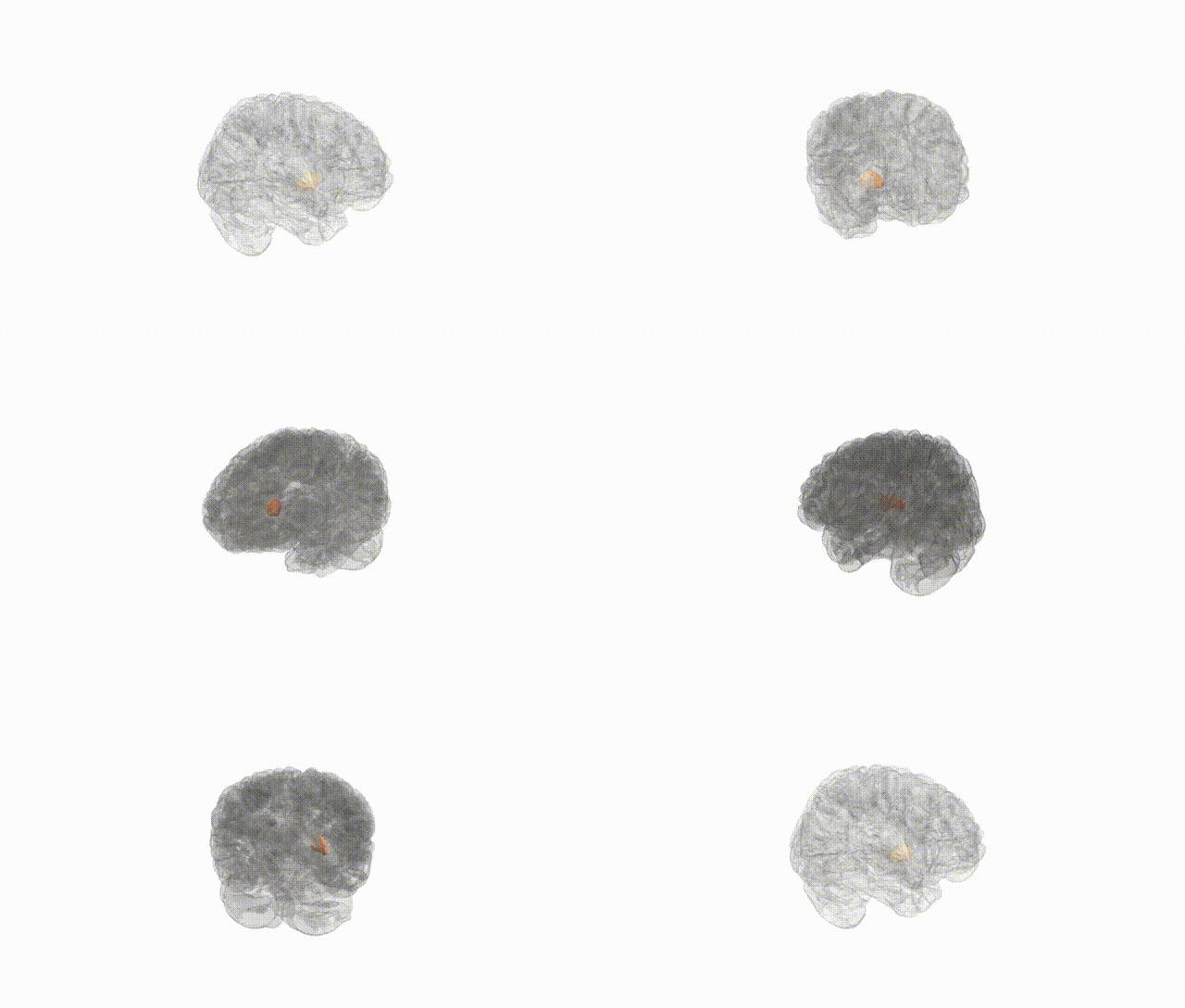
Full Quality Version: Download MP4
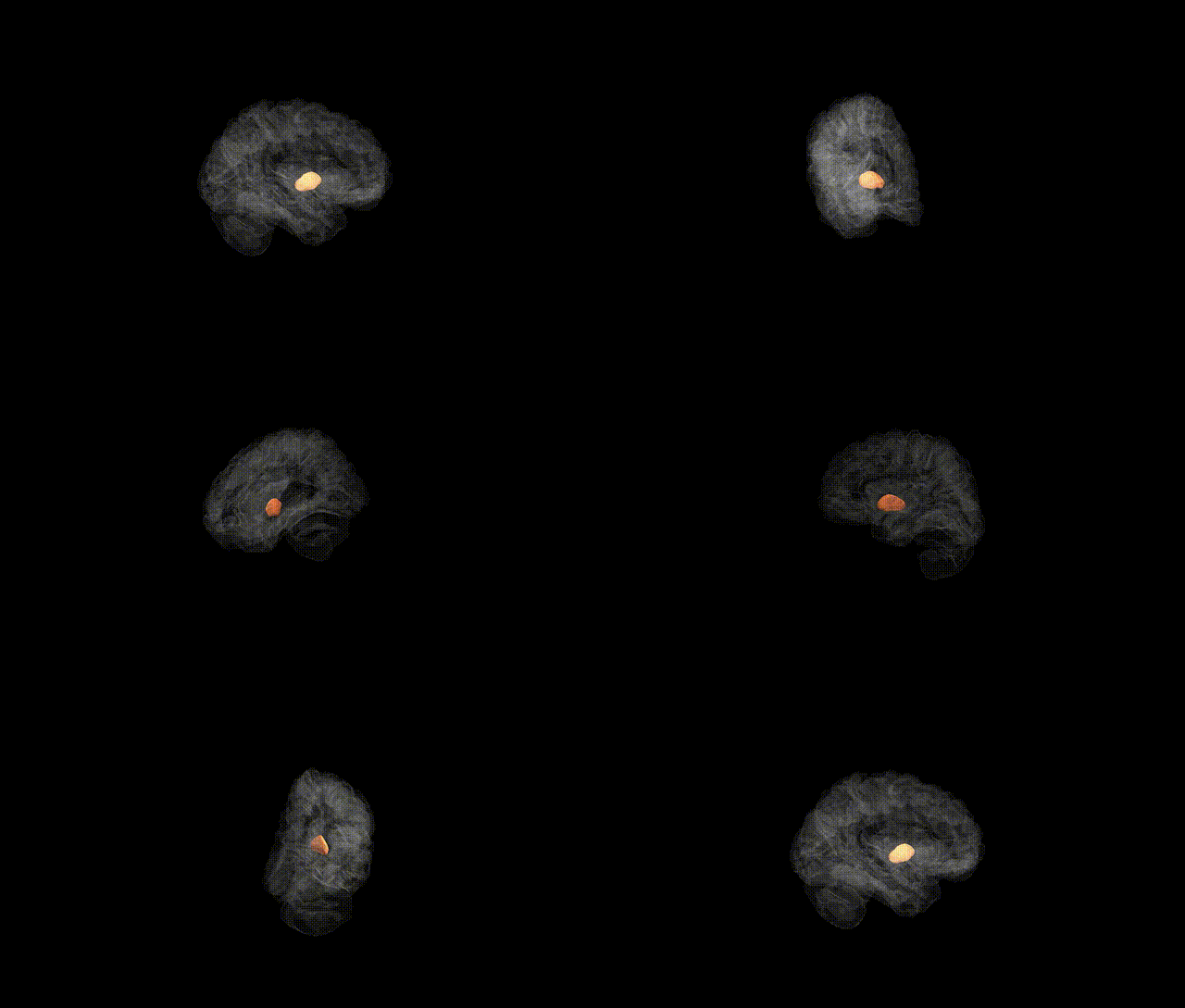
Full Quality Version: Download MP4
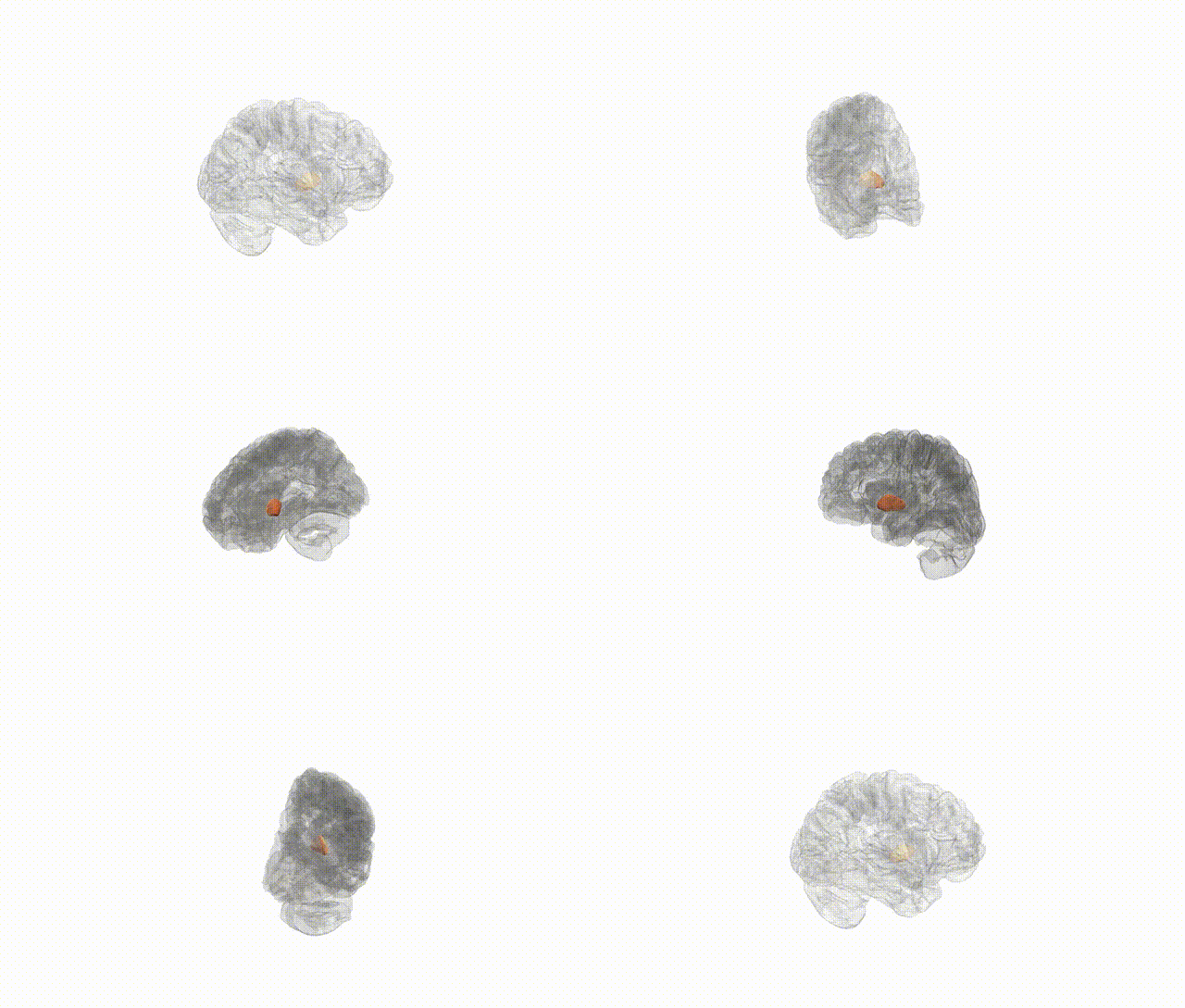
Full Quality Version: Download MP4
Wali Sidiqyar*, Gaurav Rudravaram*, Elyssa M. McMaster, Trent M. Schwartz, Adam M. Saunders, Kurt G. Schilling, Bennett A. Landman "Introducing SPINS: A Shared Public Visualization Library of Neuroanatomical Structures." Medical Imaging with Deep Learning- short paper
This resource is licensed under CC0 1.0 Universal (Public Domain).