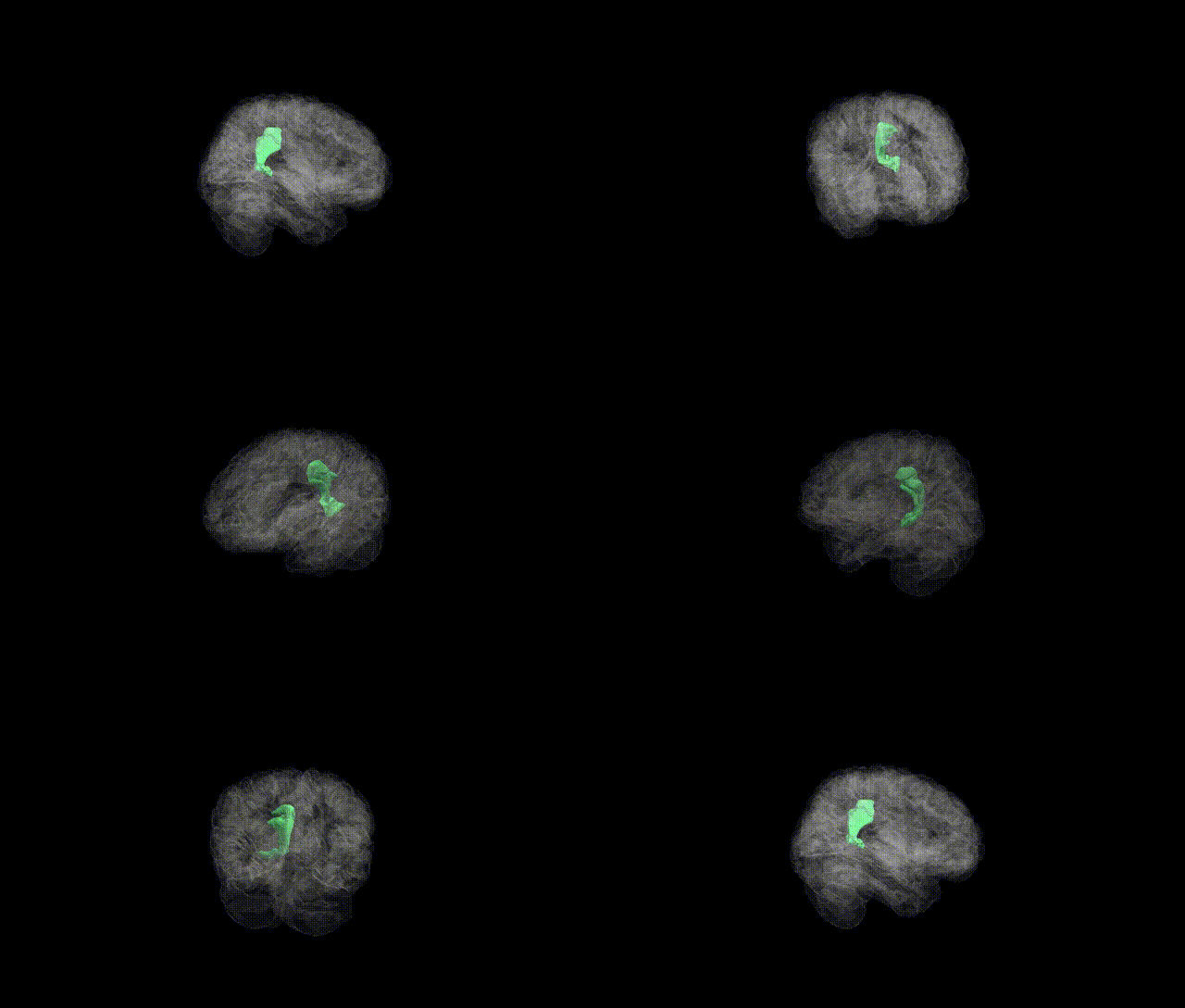
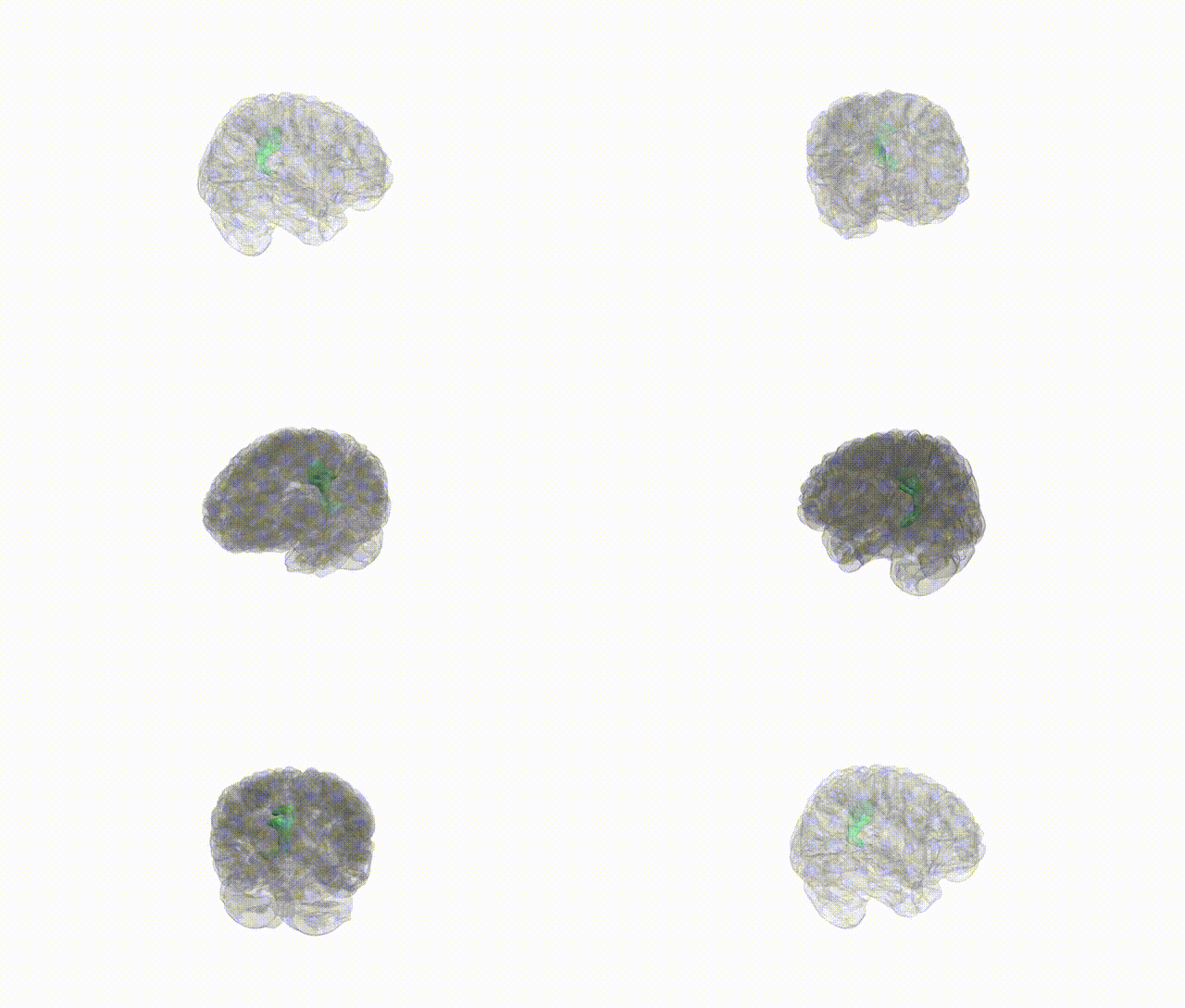
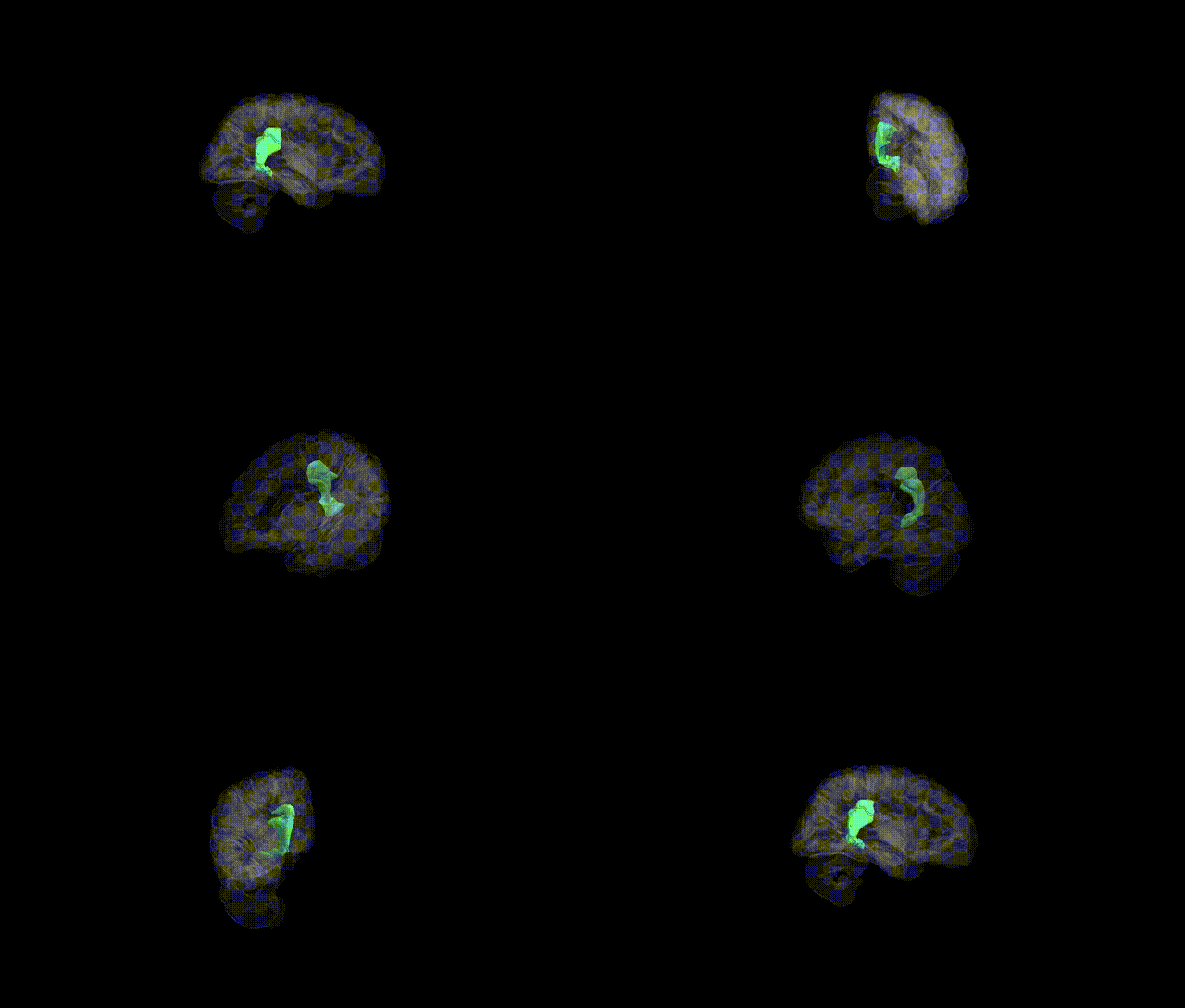
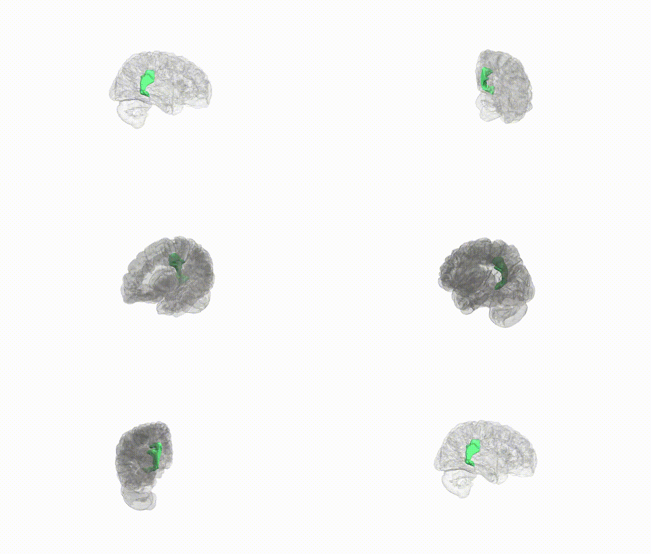
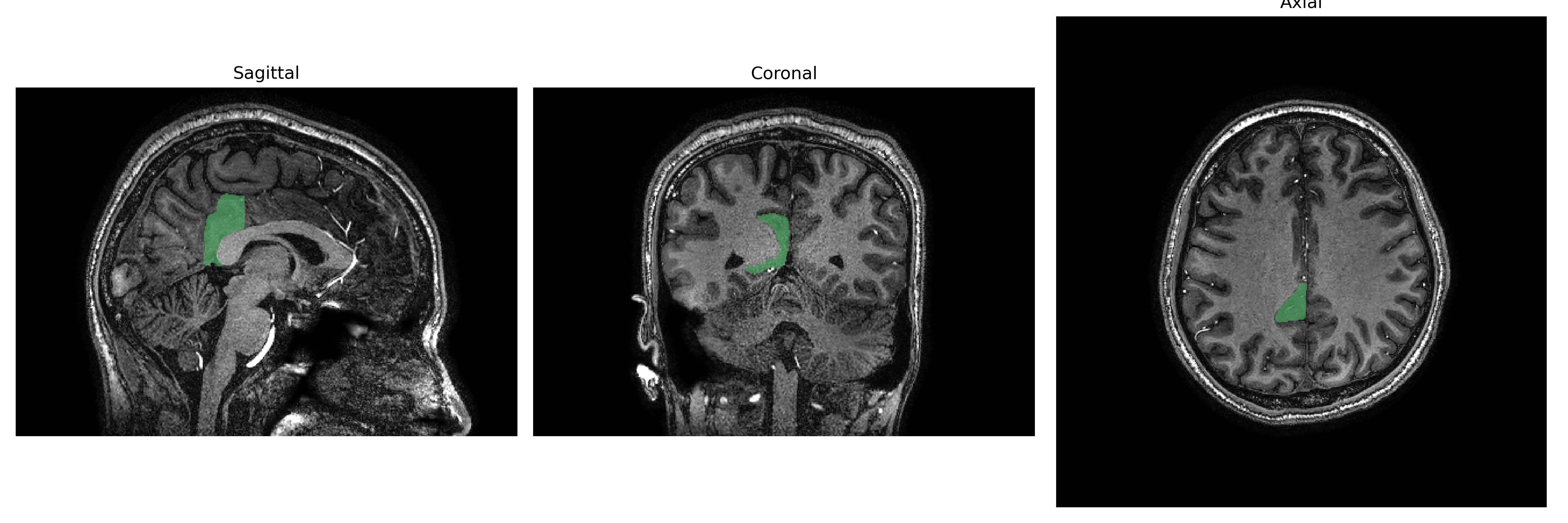
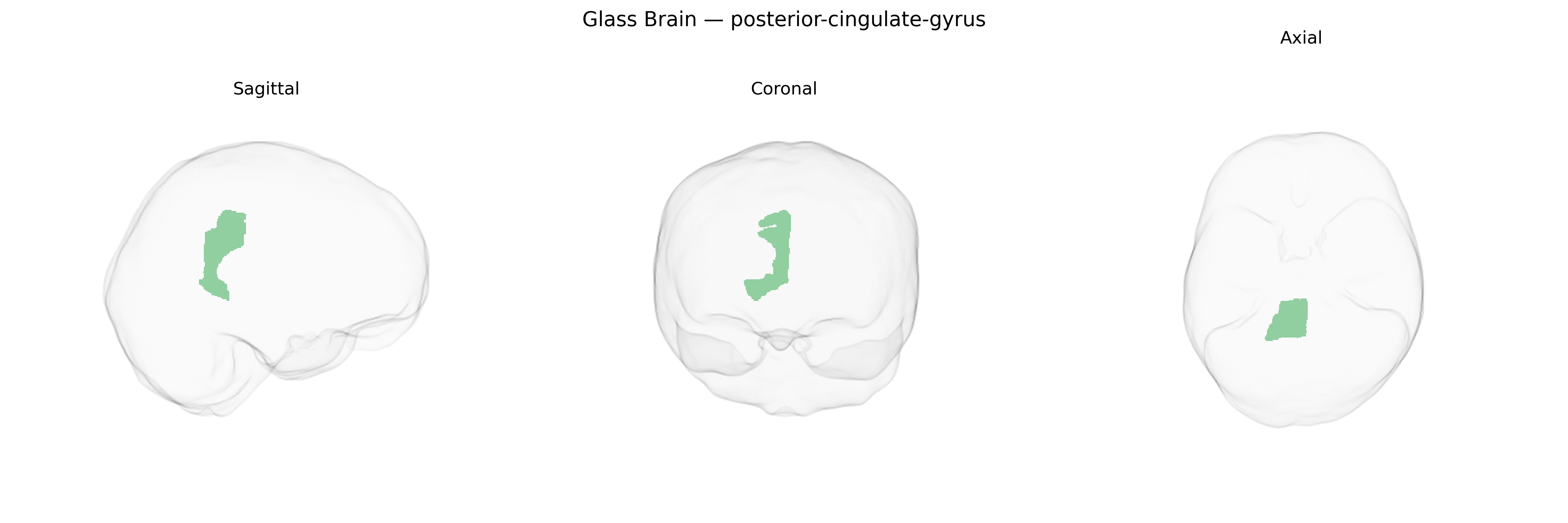
The Right posterior-cingulate-gyrus, as defined in the brainCOLOR Atlas, corresponds to the posterior segment of the cingulate gyrus located on the medial surface of the right cerebral hemisphere, bordering the precuneus and retrosplenial cortex. It forms a key hub within the default mode network and is heavily interconnected with medial prefrontal, parietal, and medial temporal regions, including the hippocampal formation. Functionally, this region is implicated in internally directed cognition, autobiographical and episodic memory retrieval, environmental orientation, and aspects of self-referential processing. It exhibits high baseline metabolic activity and is often affected early in neurodegenerative disorders such as Alzheimer’s disease, where reduced metabolism and structural changes in this region are characteristic. For general reference, see Posterior cingulate cortex.
The right posterior cingulate gyrus, as defined in the brainCOLOR Atlas, shows genetic associations primarily through GWAS of cortical thickness, surface area, and functional connectivity rather than single-gene effects, with substantial SNP-based heritability attributed to common variants distributed across the genome. Large-scale imaging–genetics consortia (e.g., ENIGMA, UK Biobank) have implicated loci near genes involved in synaptic function, neurodevelopment, and myelination (such as genes in glutamatergic signaling and axon guidance pathways) in inter-individual variation of posterior cingulate structure and connectivity, though specific lead variants and genes are often shared with broader default mode network regions rather than unique to this subregion. Polygenic risk scores for Alzheimer’s disease, major depressive disorder, schizophrenia, and autism spectrum disorder have been associated with altered structure or activity in posterior cingulate and adjacent precuneus and medial parietal areas, consistent with this region’s role in memory, self-referential processing, and network integration, and APOE-related risk for Alzheimer’s disease has repeatedly been linked to functional and metabolic alterations in posterior cingulate cortex. Additionally, GWAS of cognitive traits such as general intelligence, episodic memory, and educational attainment show enrichment of associated variants in genes expressed in posterior cingulate and default mode network hubs, suggesting that genetic influences on cognition and neuropsychiatric vulnerability partially converge on this region’s morphology and connectivity, although current evidence rarely isolates the right posterior cingulate gyrus as a uniquely or exclusively implicated structure.
Overview generated by GPT-4o (2026).
Region ID: 82
Hemisphere: Right
Atlas: brainCOLOR
Full Quality Version: Download MP4
Full Quality Version: Download MP4
Full Quality Version: Download MP4
Full Quality Version: Download MP4
Wali Sidiqyar*, Gaurav Rudravaram*, Elyssa M. McMaster, Trent M. Schwartz, Adam M. Saunders, Kurt G. Schilling, Bennett A. Landman "Introducing SPINS: A Shared Public Visualization Library of Neuroanatomical Structures." Medical Imaging with Deep Learning- short paper
This resource is licensed under CC0 1.0 Universal (Public Domain).