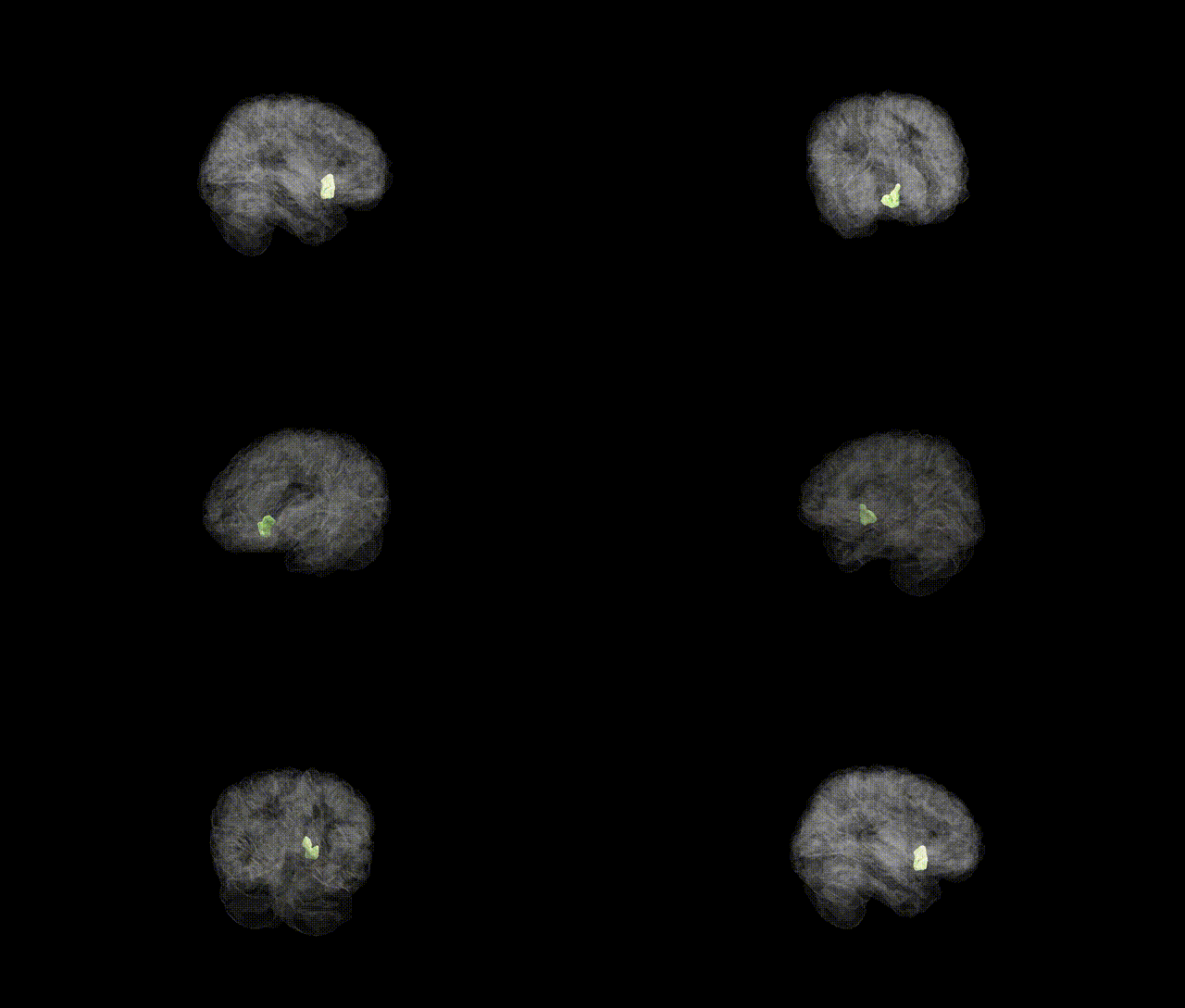
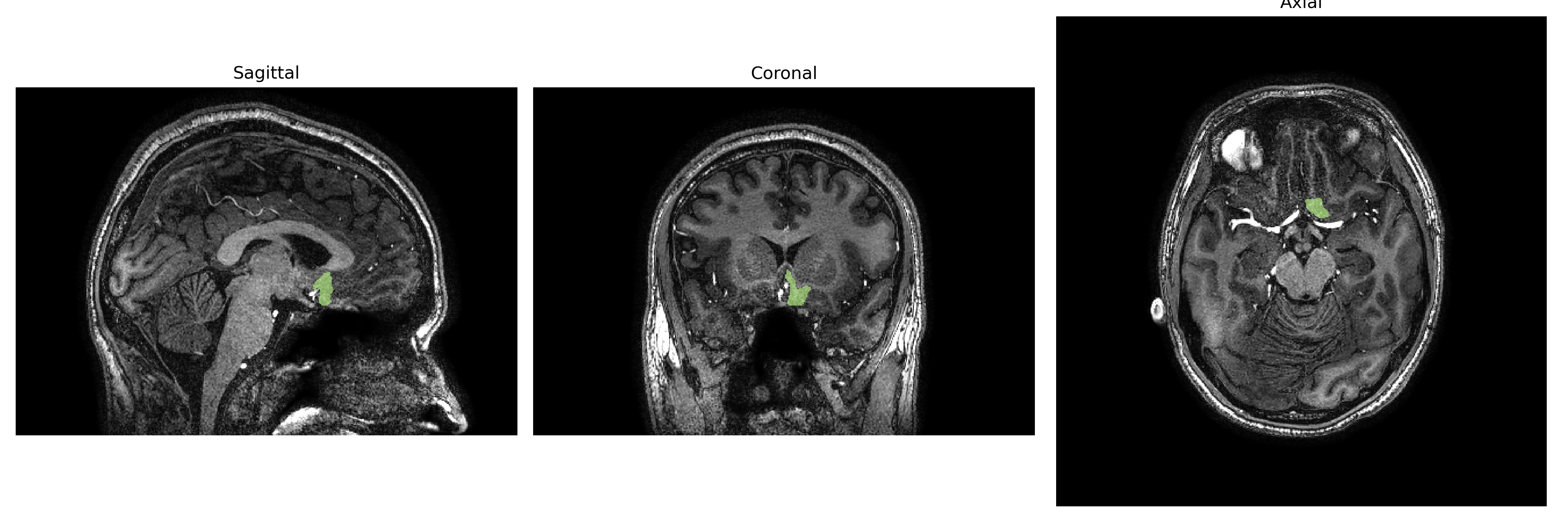
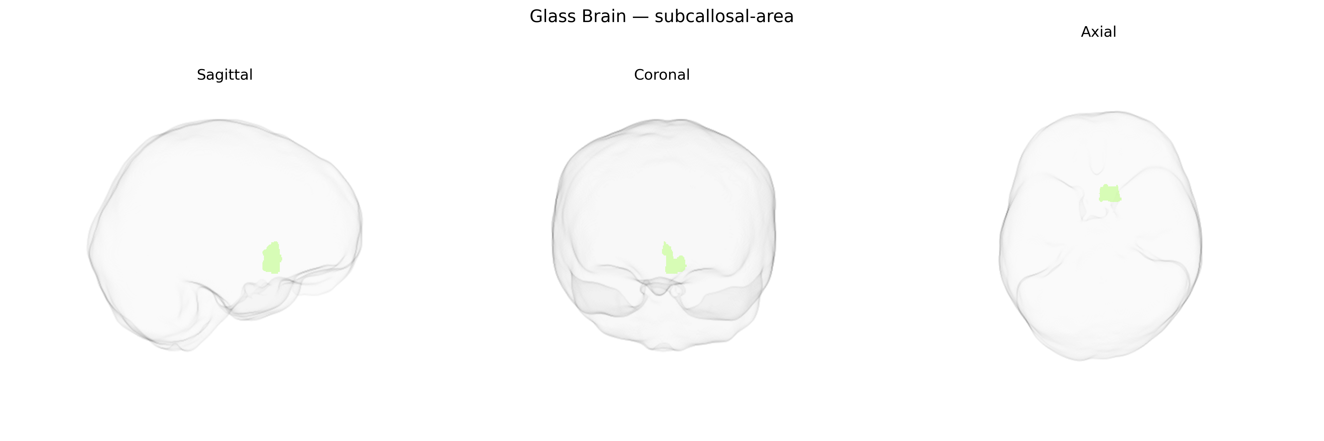
The Left subcallosal area, as defined in the brainCOLOR Atlas, is a ventromedial cortical region located beneath the rostrum of the corpus callosum within the medial frontal lobe, often considered part of the ventromedial prefrontal and limbic cortices. Cytoarchitectonically related to Brodmann area 25 (subgenual cingulate cortex), it is characterized by dense connections with the hypothalamus, amygdala, nucleus accumbens, hippocampal formation, and other cingulate and prefrontal regions, supporting roles in affective regulation, autonomic control, and visceromotor integration. This region is strongly implicated in mood disorders, particularly treatment-resistant major depression, and is a target for deep brain stimulation and other neuromodulatory interventions. There is no direct Wikipedia article specifically for the “subcallosal area”; a closely related and overlapping structure is the Subgenual cingulate cortex.
The left subcallosal area (often overlapping the subgenual anterior cingulate cortex, Brodmann area 25) has been implicated in several genetic and GWAS-based findings related to mood and affective traits, though most large-scale GWAS report associations at the level of broader anterior cingulate or limbic networks rather than this parcel alone. Variants in genes involved in monoaminergic signaling and synaptic plasticity—such as SLC6A4, BDNF (e.g., Val66Met), and genes regulating glutamatergic and GABAergic transmission—have been linked to structural and functional differences in subgenual/subcallosal regions, often in the context of major depressive disorder, bipolar disorder, and treatment response to antidepressants or deep brain stimulation. Imaging–genetics studies indicate that polygenic risk scores for depression, neuroticism, and related affective traits correlate with altered volume, cortical thickness, or activity in the subcallosal/sgACC area, and several psychiatric GWAS (including for major depressive disorder and anxiety-related traits) highlight nearby regulatory loci whose expression patterns are enriched in anterior cingulate–limbic circuits. Additionally, subcallosal area measures have been associated with variants in genes affecting white-matter development and myelination in large neuroimaging cohorts (e.g., ENIGMA, UK Biobank–based studies), though those findings typically aggregate across cingulate subregions rather than isolating the left subcallosal region as defined in the brainCOLOR Atlas.
Overview generated by GPT-4o (2026).
Region ID: 103
Hemisphere: Left
Atlas: brainCOLOR
Full Quality Version: Download MP4
Full Quality Version: Download MP4
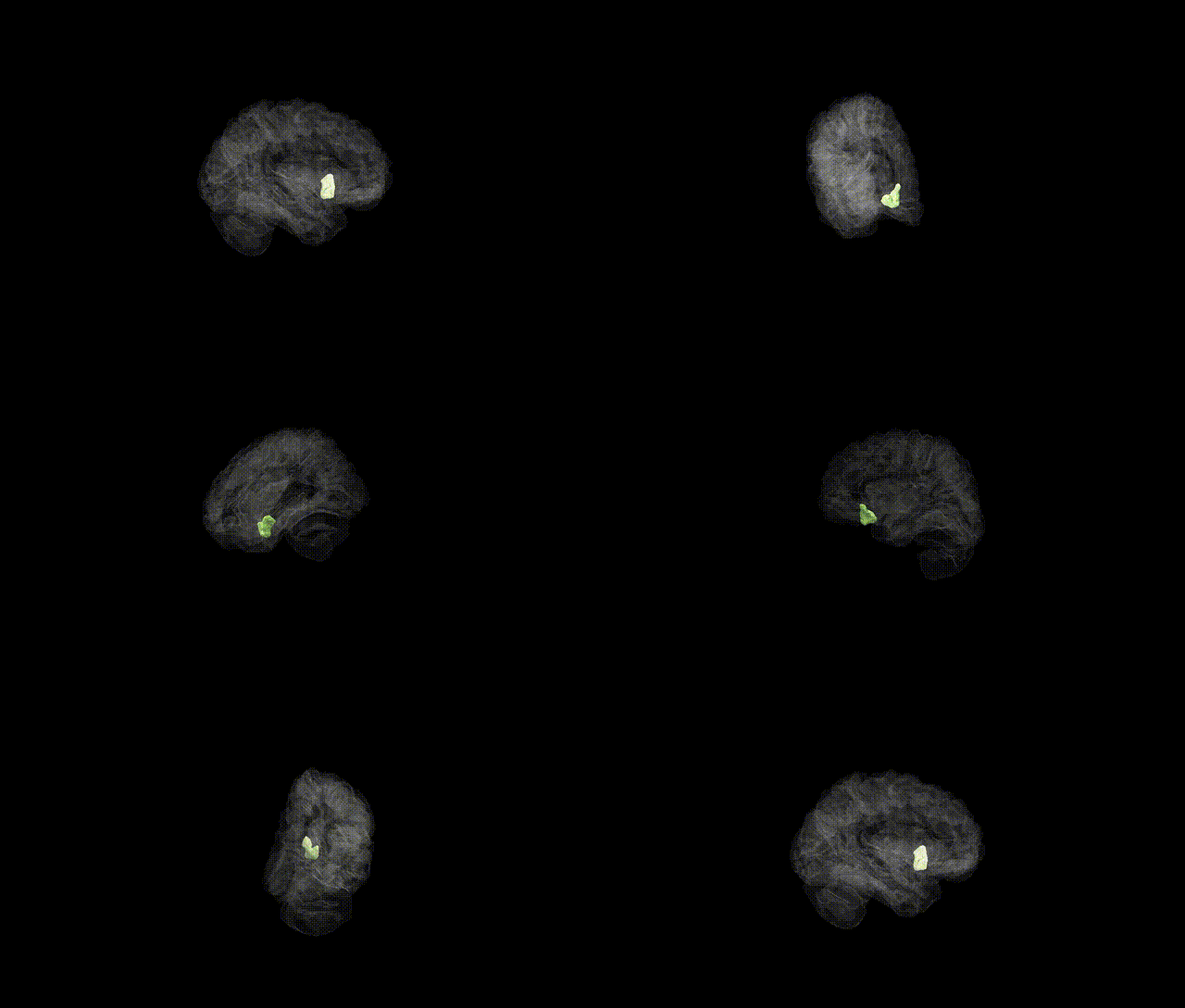
Full Quality Version: Download MP4
Full Quality Version: Download MP4
Wali Sidiqyar*, Gaurav Rudravaram*, Elyssa M. McMaster, Trent M. Schwartz, Adam M. Saunders, Kurt G. Schilling, Bennett A. Landman "Introducing SPINS: A Shared Public Visualization Library of Neuroanatomical Structures." Medical Imaging with Deep Learning- short paper
This resource is licensed under CC0 1.0 Universal (Public Domain).