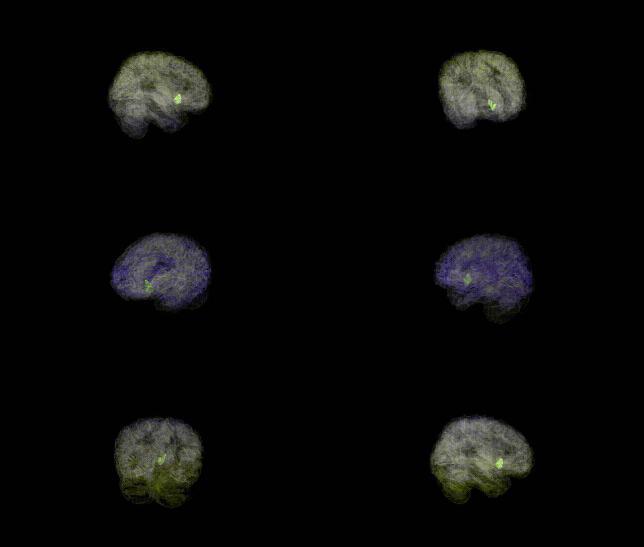
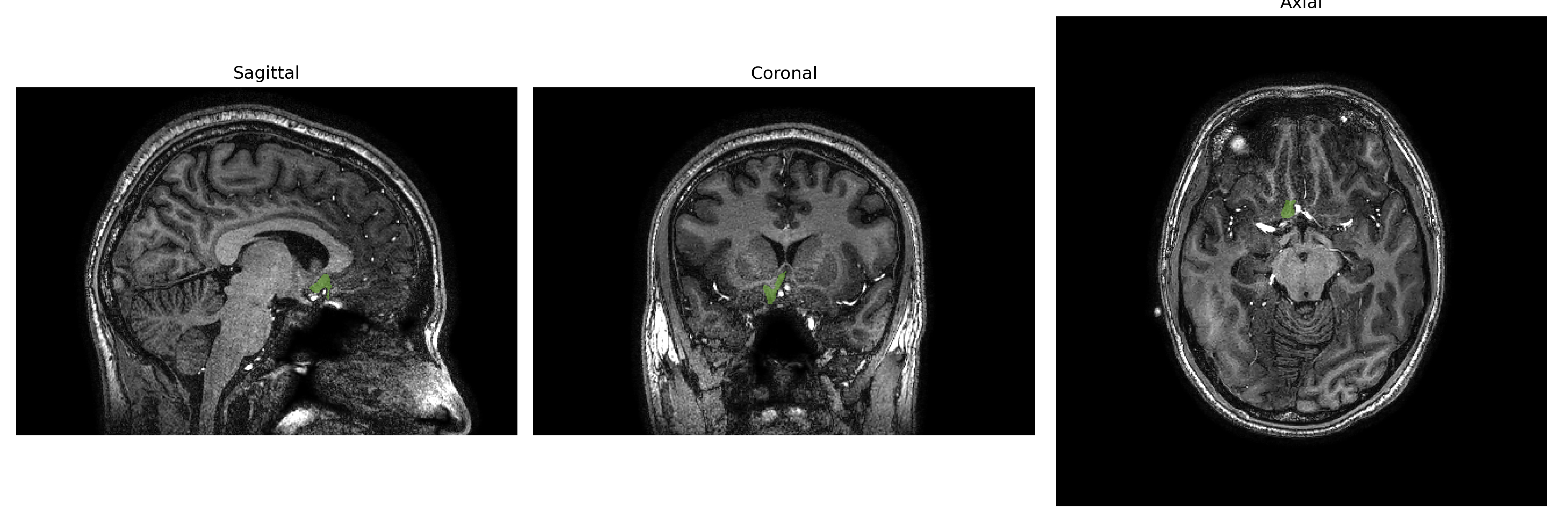
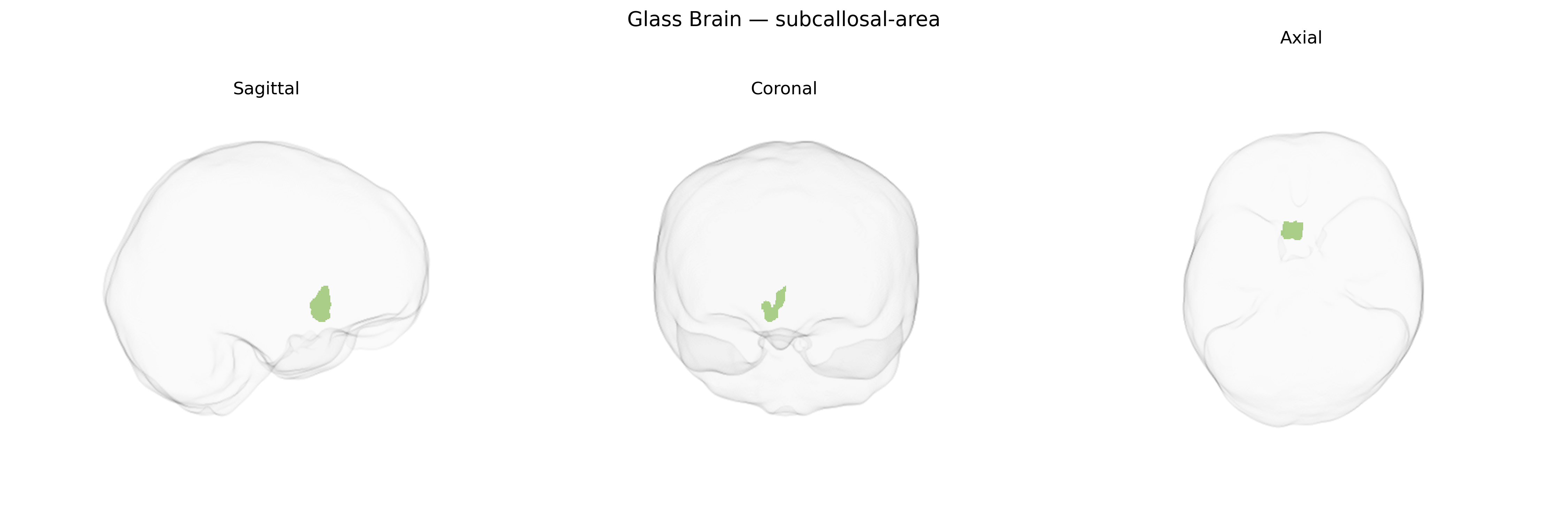
The Right subcallosal-area, as defined in the brainCOLOR Atlas, is a ventromedial frontal region located inferior to the genu of the corpus callosum, forming part of the subcallosal cortex within the medial prefrontal and limbic network. It encompasses cortex and adjacent white matter around the subcallosal gyrus and is closely associated with the subgenual anterior cingulate cortex (often corresponding to Brodmann area 25). This region is heavily interconnected with limbic structures such as the amygdala, hypothalamus, and ventral striatum, and receives dense monoaminergic input, particularly serotonergic and dopaminergic projections. Functionally, it is implicated in mood regulation, autonomic and visceromotor control, and affective processing, and has been a key target in neuromodulatory interventions (e.g., deep brain stimulation) for treatment-resistant major depressive disorder.
The right subcallosal area (subcallosal cortex/subgenual anterior cingulate region) from the brainCOLOR atlas has been implicated in several genetic and GWAS-based findings, largely through studies of subcortical volume, cortical thickness, and functional/anatomical connectivity. Large neuroimaging genetics consortia such as ENIGMA and UK Biobank–based GWAS have reported associations between common variants in genes involved in neurodevelopment, synaptic plasticity, and axon guidance (for example, near genes such as CACNA1C, TCF4, and those within the major histocompatibility complex) and structural variation in ventral medial prefrontal and cingulate regions encompassing the subcallosal area. This region shows robust heritability of volume and thickness and emerges in polygenic risk analyses linking higher genetic liability for major depressive disorder, bipolar disorder, and schizophrenia to altered subcallosal/ventromedial prefrontal morphology and activity. Variants in serotonin and glutamate-related genes (e.g., SLC6A4, GRM family members) and depression- and mood-related polygenic scores have been tied to functional changes and connectivity patterns involving the subcallosal area, consistent with its role as a key node in mood regulation networks and as a target of deep brain stimulation for treatment-resistant depression. Overall, GWAS and candidate-gene imaging genetics converge in indicating that common genetic variation influencing psychiatric risk, affective traits, and stress sensitivity is associated with structural and functional variation in the right subcallosal area.
Overview generated by GPT-4o (2026).
Region ID: 102
Hemisphere: Right
Atlas: brainCOLOR
Full Quality Version: Download MP4
Full Quality Version: Download MP4
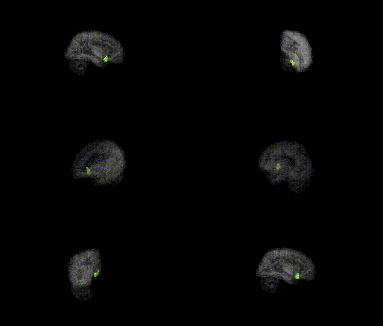
Full Quality Version: Download MP4
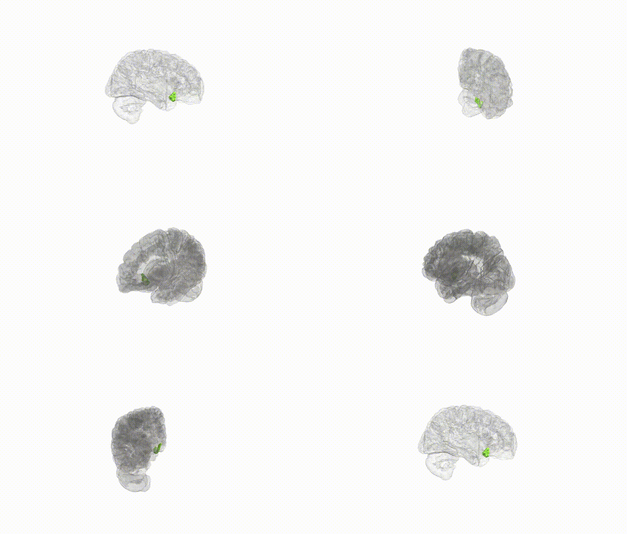
Full Quality Version: Download MP4
Wali Sidiqyar*, Gaurav Rudravaram*, Elyssa M. McMaster, Trent M. Schwartz, Adam M. Saunders, Kurt G. Schilling, Bennett A. Landman "Introducing SPINS: A Shared Public Visualization Library of Neuroanatomical Structures." Medical Imaging with Deep Learning- short paper
This resource is licensed under CC0 1.0 Universal (Public Domain).