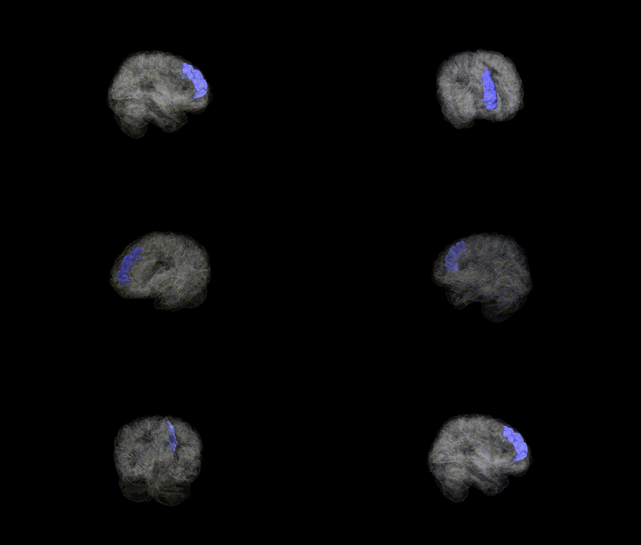
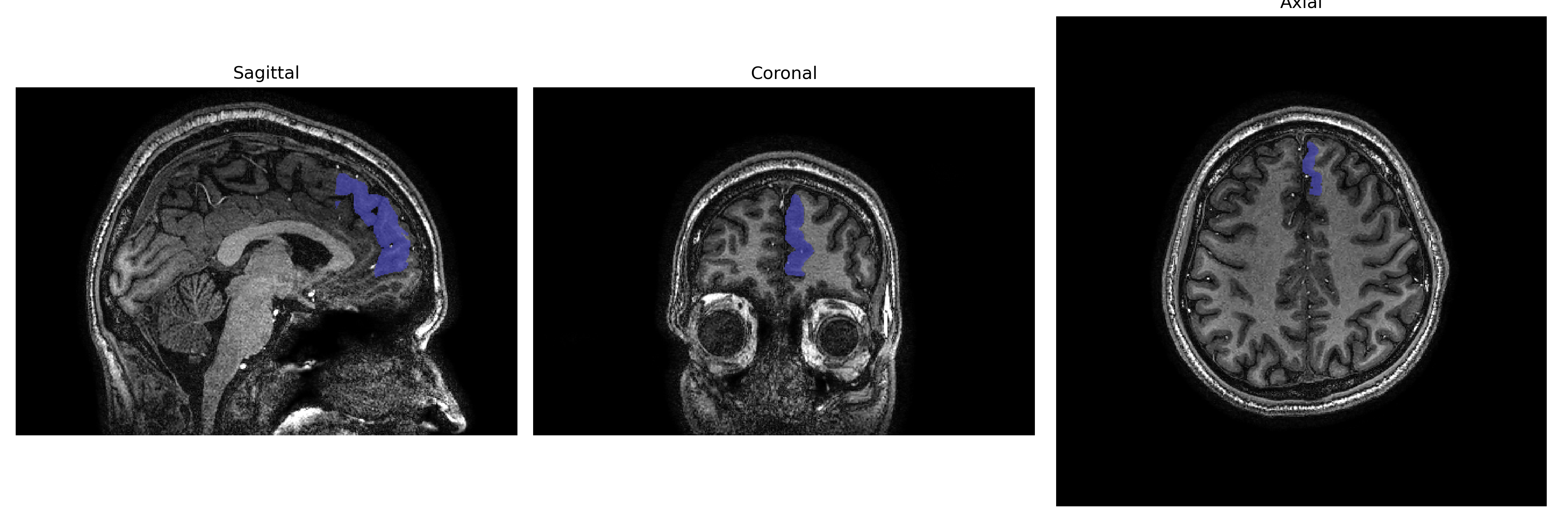
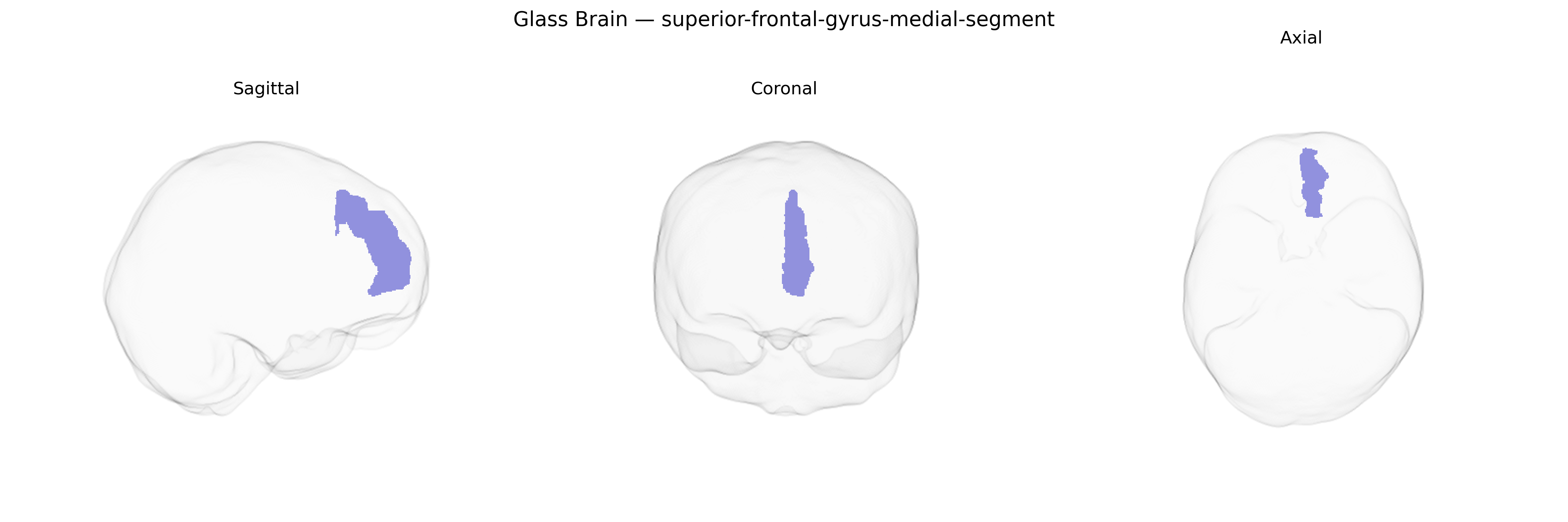
The left superior frontal gyrus, medial segment, corresponds to the medial portion of the superior frontal gyrus within the dorsomedial prefrontal cortex on the medial surface of the left frontal lobe. It is situated anterior to the supplementary motor area and superior to the cingulate sulcus, participating in high-level executive functions, self-referential processing, decision-making, and aspects of working memory and cognitive control. This region shows strong connectivity with other prefrontal, cingulate, and parietal areas and is often implicated in networks subserving attention, introspection, and regulation of goal-directed behavior. There is no direct Wikipedia article specifically for the “medial segment” of the left superior frontal gyrus; a related general entry is Superior frontal gyrus.
The left superior frontal gyrus (medial segment), as defined in parcellations such as the brainCOLOR Atlas, has been implicated in several genetic and GWAS-based findings through its roles in executive control, self-referential processing, and mood regulation. Large-scale imaging genetics consortia (e.g., ENIGMA, UK Biobank–based GWAS of cortical thickness and surface area) have identified common variants in genes involved in neurodevelopment, synaptic signaling, and cell adhesion (including loci near MIR137, CNTNAP2, CACNA1C, and genes in the major histocompatibility complex region) that show associations with structural variation in medial superior frontal regions, encompassing the medial segment of the superior frontal gyrus. Polygenic risk scores for schizophrenia, major depressive disorder, bipolar disorder, attention-deficit/hyperactivity disorder, and autism spectrum disorder have been linked to altered cortical thickness, surface area, or functional connectivity in medial superior frontal cortex, with some studies showing that greater genetic liability to these disorders corresponds to volumetric or functional changes in this region. GWAS of cognitive traits (general intelligence, educational attainment, and executive function) and personality dimensions (notably neuroticism and conscientiousness) also report that polygenic scores for these traits correlate with structural and functional metrics in medial superior frontal areas. In addition, risk variants for mood and anxiety disorders and for internalizing psychopathology more broadly have been associated with altered resting-state connectivity of the medial superior frontal gyrus within the default mode and frontoparietal control networks, although specific variant–region links are typically modest in effect size and distributed, reflecting the highly polygenic architecture of these brain phenotypes.
Overview generated by GPT-4o (2026).
Region ID: 71
Hemisphere: Left
Atlas: brainCOLOR
Full Quality Version: Download MP4
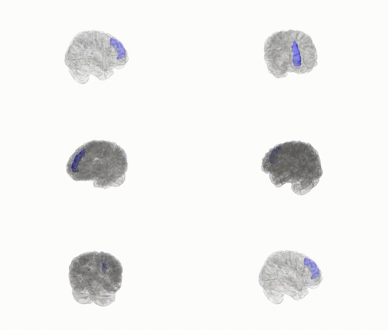
Full Quality Version: Download MP4
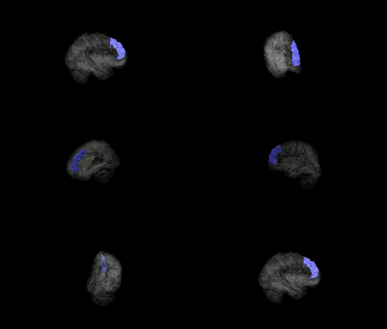
Full Quality Version: Download MP4
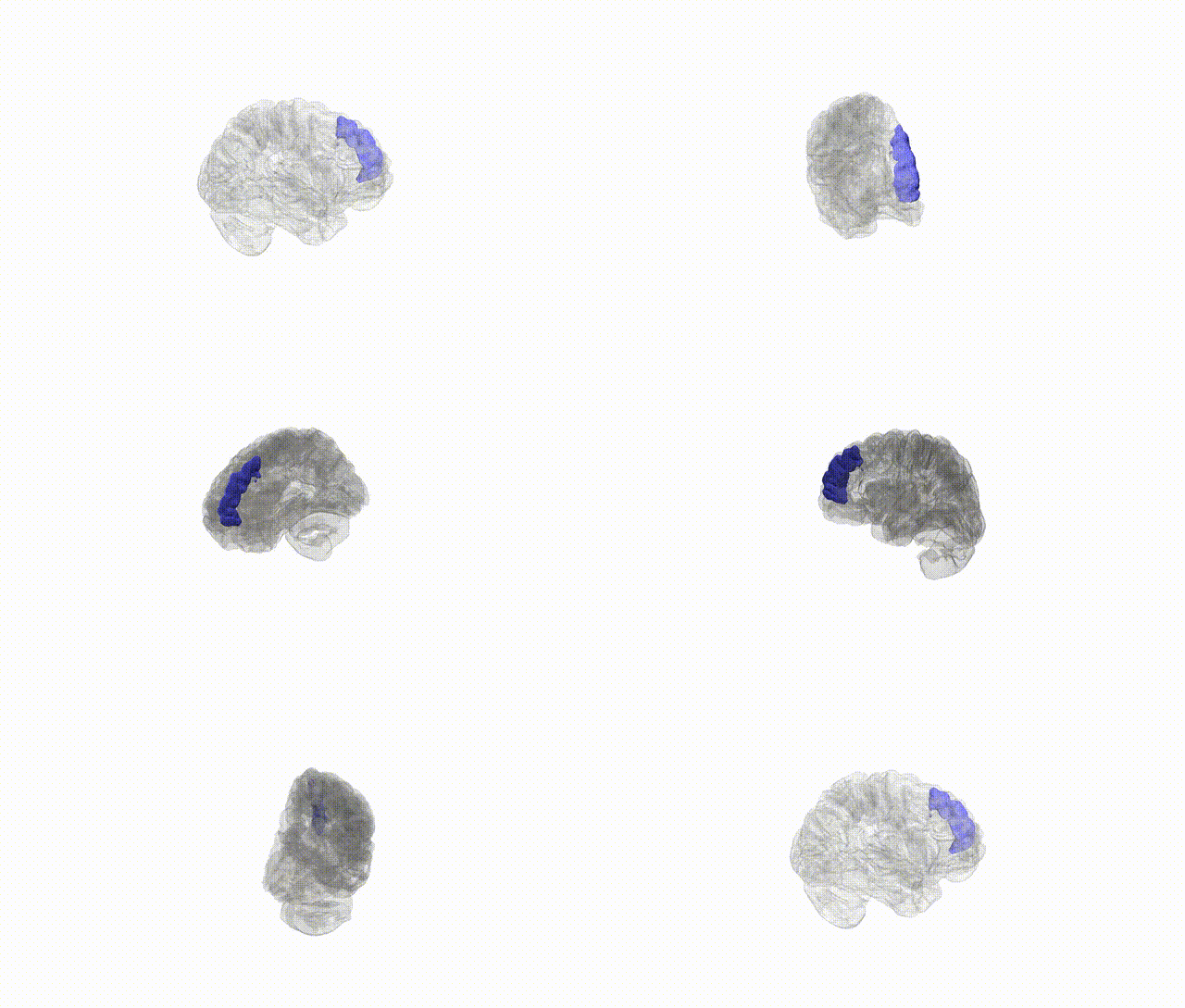
Full Quality Version: Download MP4
Wali Sidiqyar*, Gaurav Rudravaram*, Elyssa M. McMaster, Trent M. Schwartz, Adam M. Saunders, Kurt G. Schilling, Bennett A. Landman "Introducing SPINS: A Shared Public Visualization Library of Neuroanatomical Structures." Medical Imaging with Deep Learning- short paper
This resource is licensed under CC0 1.0 Universal (Public Domain).