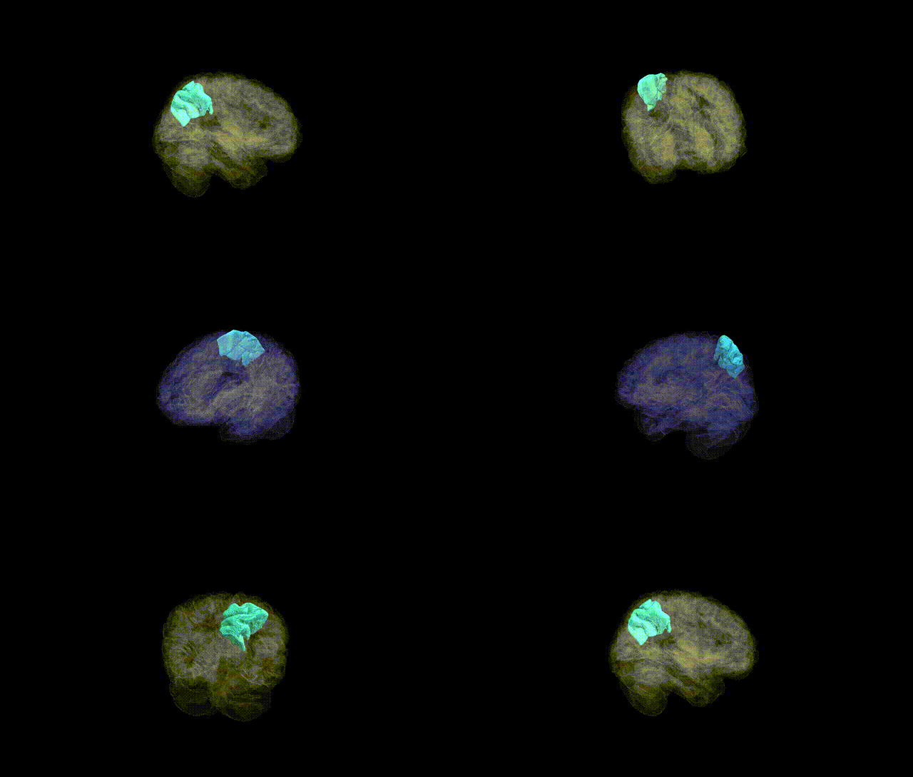
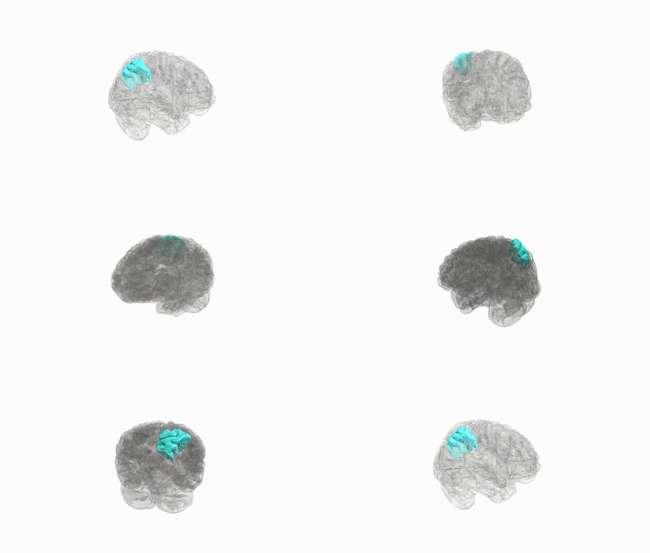
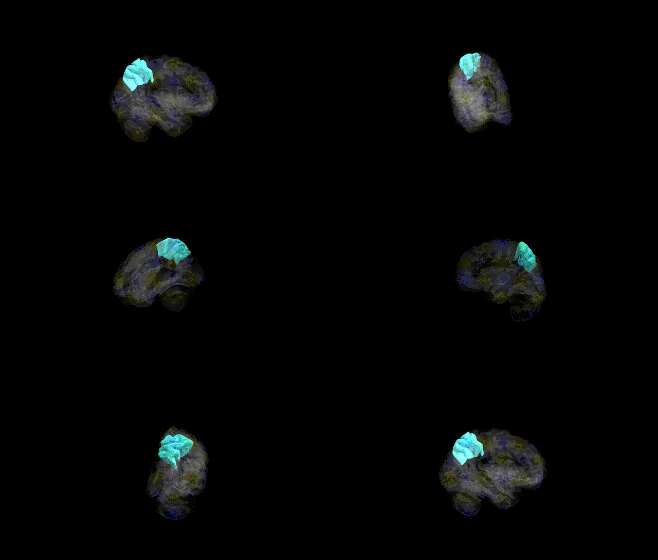
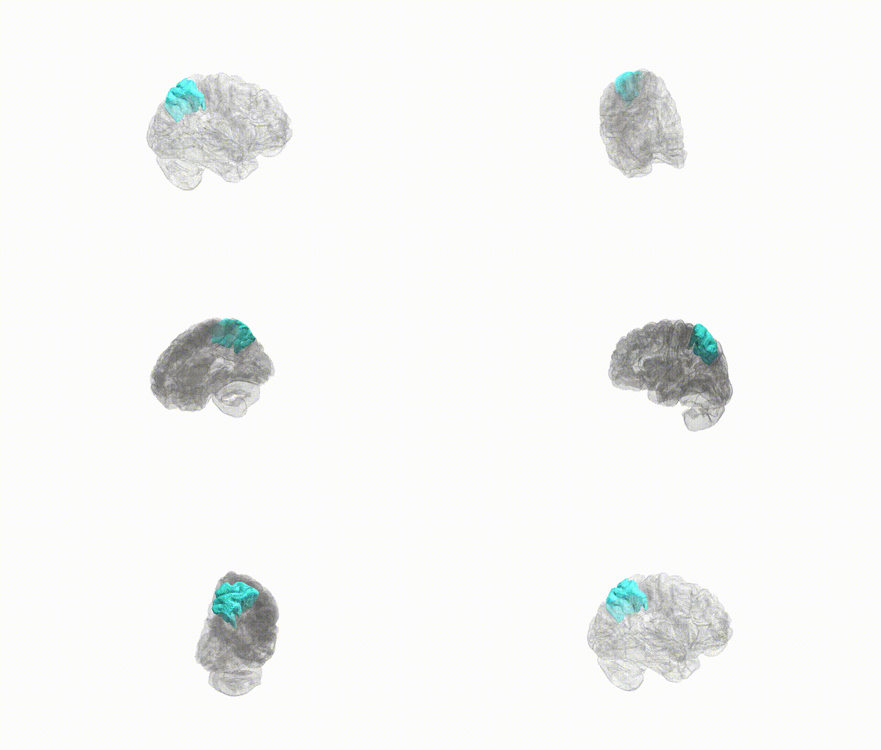
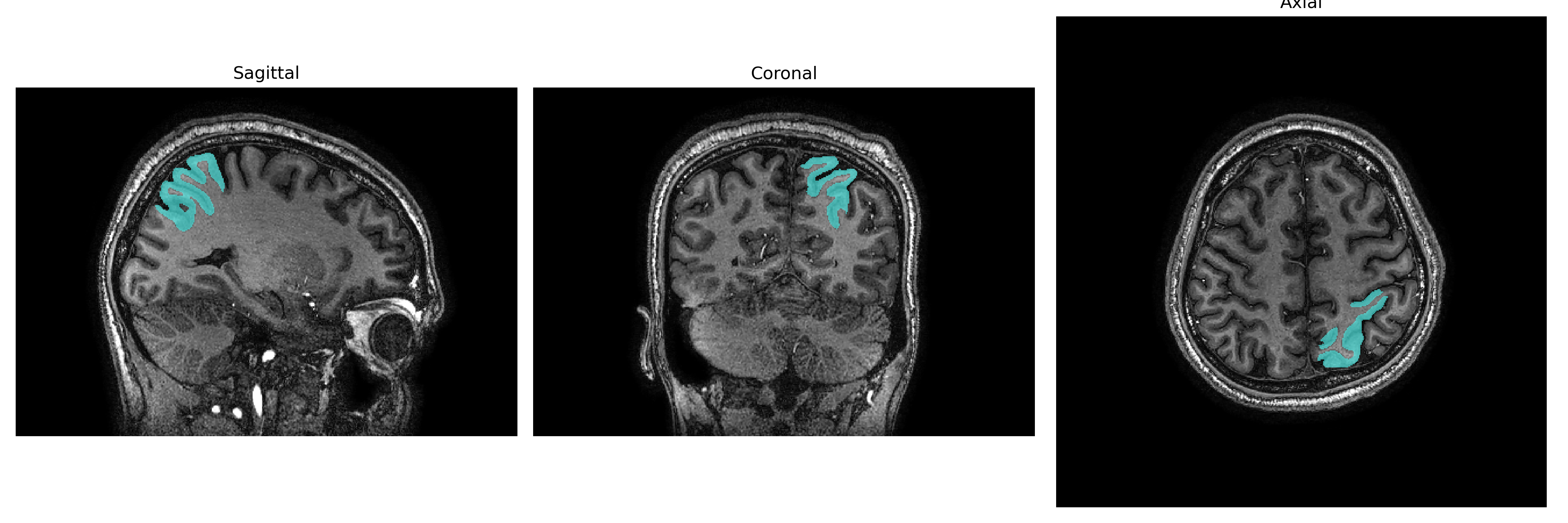
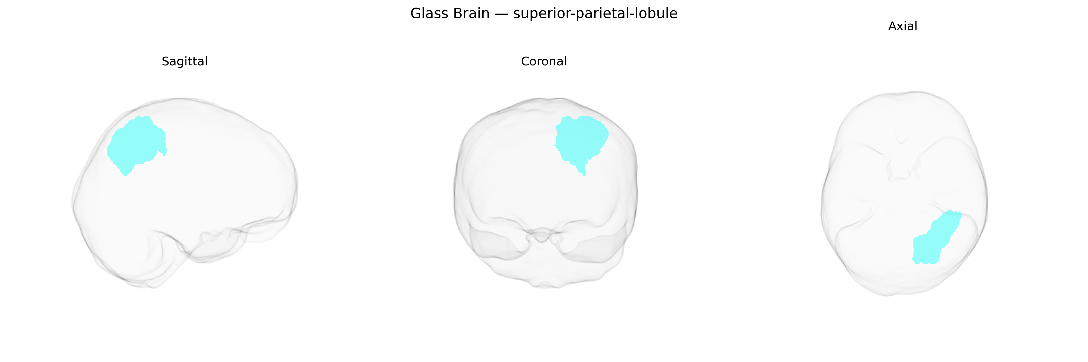
The Left superior parietal lobule is a dorsal parietal cortex region located on the lateral surface of the left hemisphere, bounded anteriorly by the postcentral gyrus, medially by the paracentral lobule and precuneus, and inferiorly by the intraparietal sulcus separating it from the inferior parietal lobule. Cytoarchitectonically, it encompasses portions of Brodmann areas 5 and 7, with dense reciprocal connections to primary and secondary somatosensory cortices, premotor areas, occipital visual regions, and subcortical structures such as the thalamus. Functionally, it is critically involved in multisensory integration, visuospatial processing, body schema representation, and the guidance of goal-directed actions (e.g., reaching and grasping), and it contributes to attentional control and spatial working memory. Lesions in this region, particularly in the dominant hemisphere, can lead to deficits such as optic ataxia, impaired spatial orientation, and components of hemispatial neglect or apraxia.
Superior parietal lobule
The left superior parietal lobule (SPL), as defined in the brainCOLOR atlas, has been implicated in several genetic and GWAS findings related to cortical structure, cognition, and neuropsychiatric risk. Large-scale imaging-genetics consortia such as ENIGMA and UK Biobank have identified common variants in genes involved in neurodevelopment, synaptic function, and axon guidance (for example, loci near genes such as HMGA2, IGF1, and various cell-adhesion and transcription-regulation genes) that are associated with SPL cortical thickness, surface area, or overall parietal lobe morphology. Polygenic scores for general cognitive ability and educational attainment are positively associated with structural and functional measures in the superior parietal region, reflecting its role in attention, working memory, and visuospatial processing. In addition, risk variants for disorders including schizophrenia, autism spectrum disorder, attention-deficit/hyperactivity disorder, and Alzheimer’s disease show convergent effects on SPL structure or connectivity, with some GWAS signals for these conditions mapping to genes that are preferentially expressed in parietal association cortex or influence synaptic plasticity and cortical patterning. Although many findings remain regionally coarse and not always specific to the left SPL parcel in the brainCOLOR atlas, converging genetic evidence indicates that this region is a key target of polygenic influences on higher-order cognition and vulnerability to major neuropsychiatric and neurodegenerative disorders.
Overview generated by GPT-4o (2026).
Region ID: 113
Hemisphere: Left
Atlas: brainCOLOR
Full Quality Version: Download MP4
Full Quality Version: Download MP4
Full Quality Version: Download MP4
Full Quality Version: Download MP4
Wali Sidiqyar*, Gaurav Rudravaram*, Elyssa M. McMaster, Trent M. Schwartz, Adam M. Saunders, Kurt G. Schilling, Bennett A. Landman "Introducing SPINS: A Shared Public Visualization Library of Neuroanatomical Structures." Medical Imaging with Deep Learning- short paper
This resource is licensed under CC0 1.0 Universal (Public Domain).