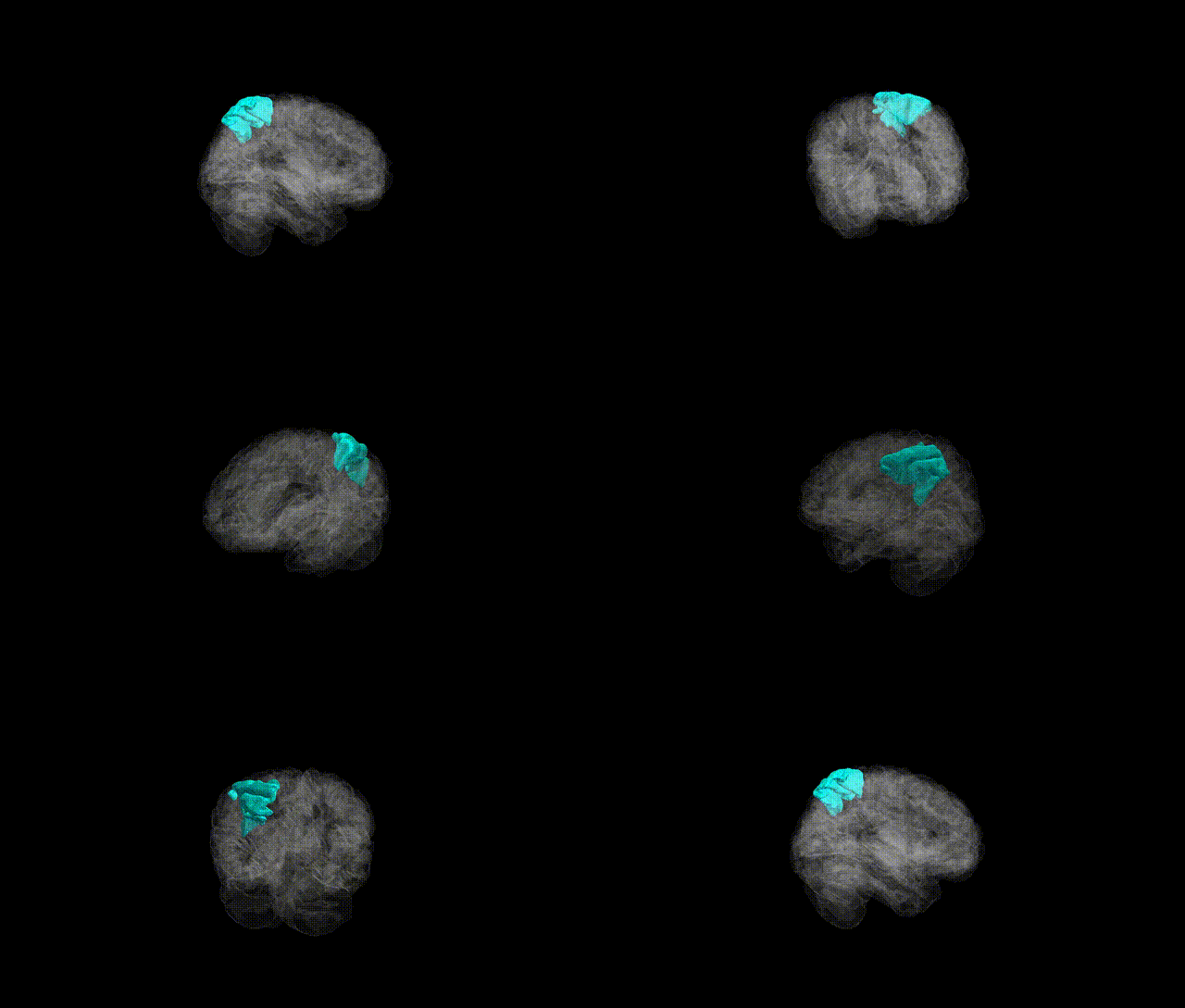
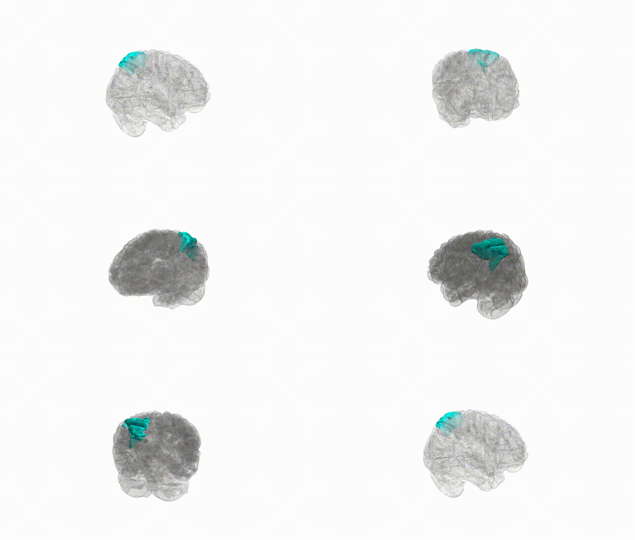
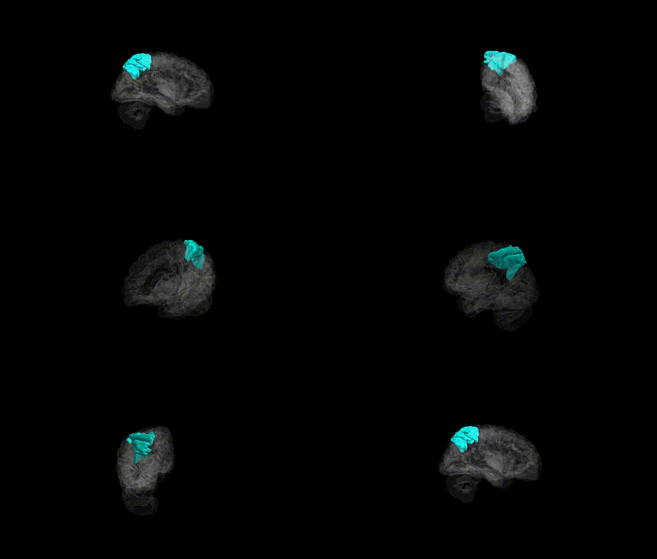
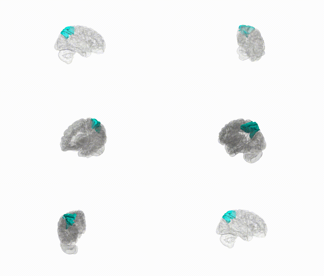
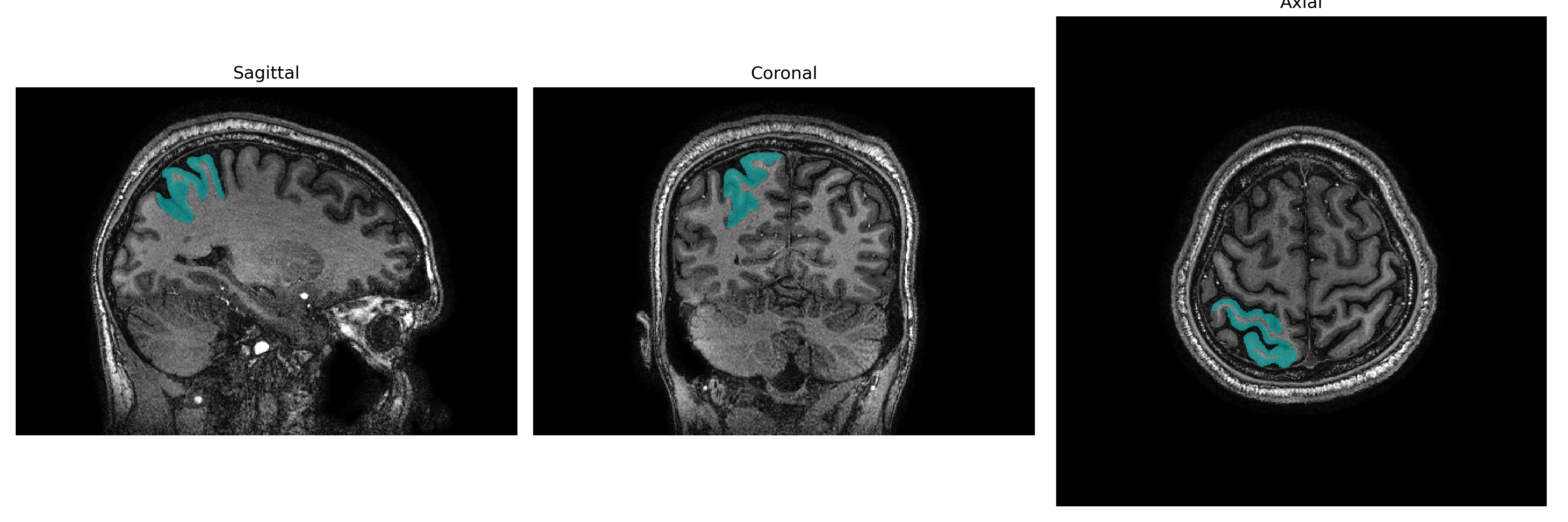
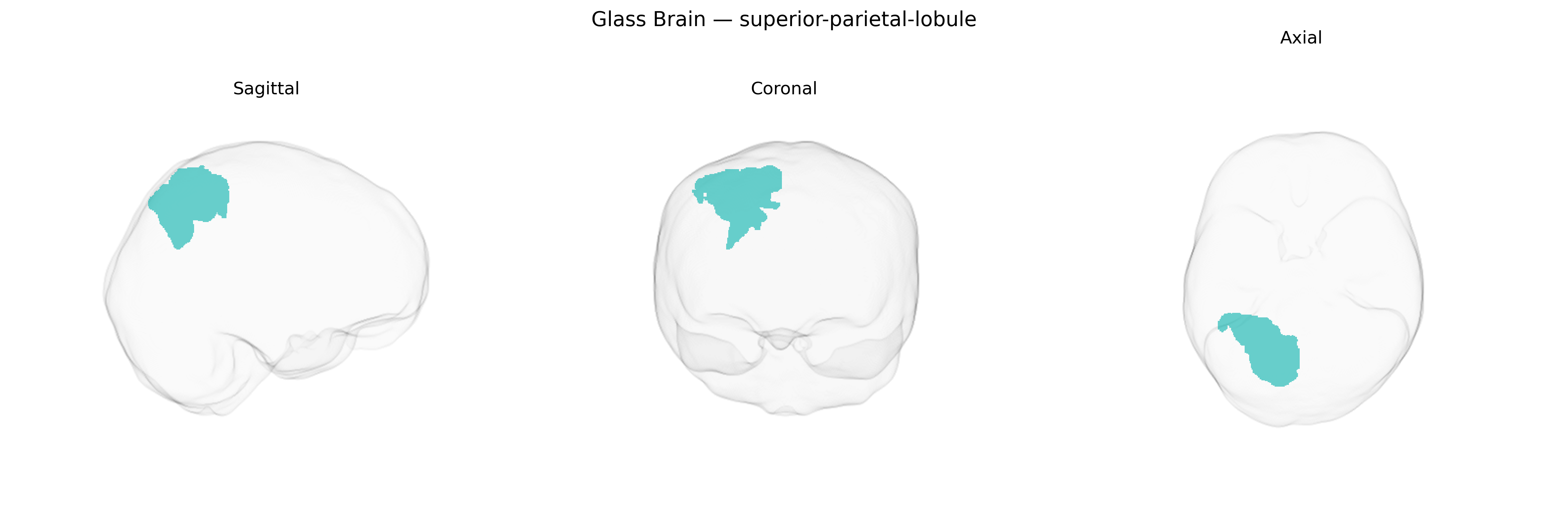
The Right superior-parietal-lobule is a dorsal parietal association region located posterior to the postcentral gyrus and superior to the intraparietal sulcus in the right hemisphere, forming part of the superior parietal lobule (SPL). Cytoarchitectonically, it includes portions of Brodmann areas 5 and 7, which integrate multimodal sensory inputs, particularly somatosensory and visual information, to support spatial perception, sensorimotor transformation, and the guidance of limb movements in extrapersonal space. This region is implicated in attention shifting, visuospatial working memory, body schema representation, and coordination of eye–hand movements, with right-hemisphere specializations often related to spatial awareness and neglect syndromes when damaged. Through extensive connectivity with premotor, frontal eye field, occipital visual, and inferior parietal regions, the Right superior-parietal-lobule contributes to higher-order visuomotor control and goal-directed behavior. There is no direct link for the right-sided subdivision; see the related structure: Superior parietal lobule.
The right superior parietal lobule (SPL), as defined in the brainCOLOR atlas, has been implicated in several genetic and genome-wide association studies (GWAS) focused on cortical structure, cognition, and neuropsychiatric risk. Large imaging-genetics consortia (e.g., ENIGMA, UK Biobank) have identified common variants near genes involved in neurodevelopment, synaptic function, and cytoskeletal organization (such as HMGA2, IGF1, and variants in 3p24–3p26 and 15q regions) that associate with SPL cortical thickness and surface area, although effects are typically small and polygenic. Right SPL morphology shows heritability estimates in the moderate range and contributes to multivariate polygenic scores for general cognitive ability, visuospatial processing, and attentional control. Genetic risk for neurodevelopmental and psychiatric conditions—including autism spectrum disorder, attention-deficit/hyperactivity disorder, and schizophrenia—has been associated with altered structure or activation in the right SPL, with polygenic risk scores for these disorders correlating with parietal measures in some cohorts. Additional GWAS link parietal structural variation, including the right SPL, to traits such as educational attainment, intelligence, and risk-taking, suggesting that this region is a convergent anatomical substrate through which broadly distributed genetic effects on cognition and behavior are partially expressed.
Overview generated by GPT-4o (2026).
Region ID: 112
Hemisphere: Right
Atlas: brainCOLOR
Full Quality Version: Download MP4
Full Quality Version: Download MP4
Full Quality Version: Download MP4
Full Quality Version: Download MP4
Wali Sidiqyar*, Gaurav Rudravaram*, Elyssa M. McMaster, Trent M. Schwartz, Adam M. Saunders, Kurt G. Schilling, Bennett A. Landman "Introducing SPINS: A Shared Public Visualization Library of Neuroanatomical Structures." Medical Imaging with Deep Learning- short paper
This resource is licensed under CC0 1.0 Universal (Public Domain).