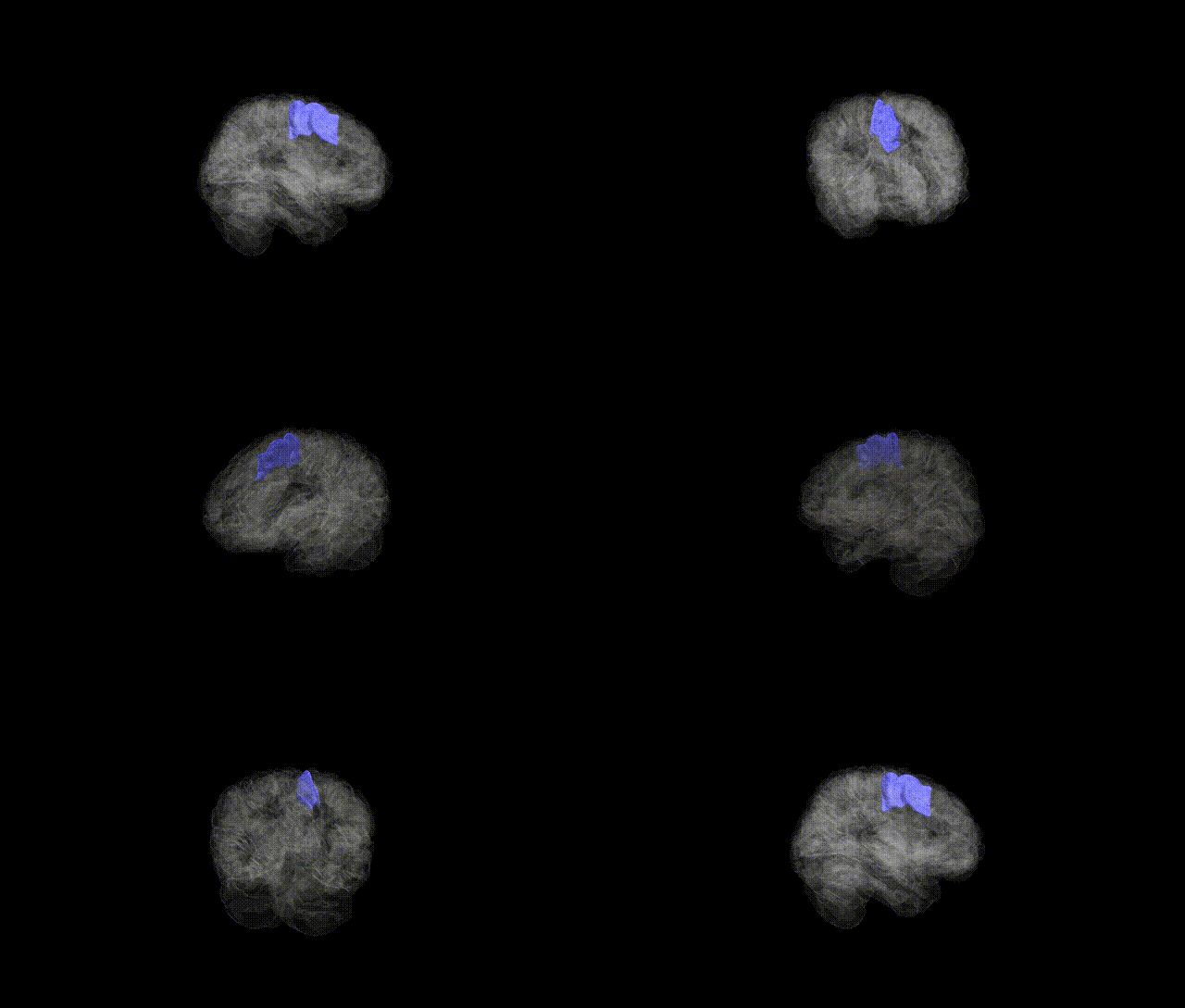
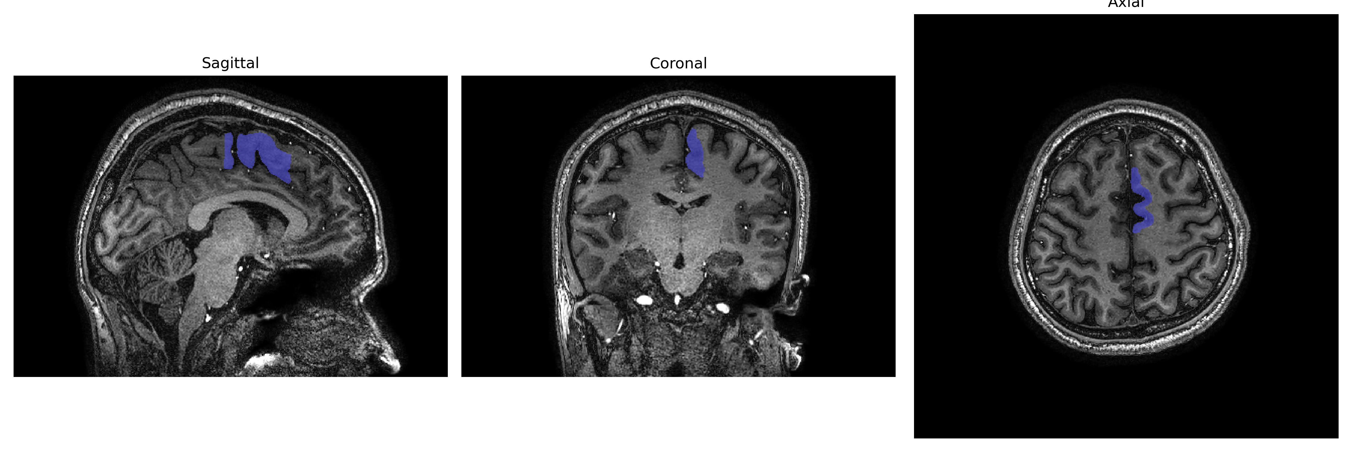
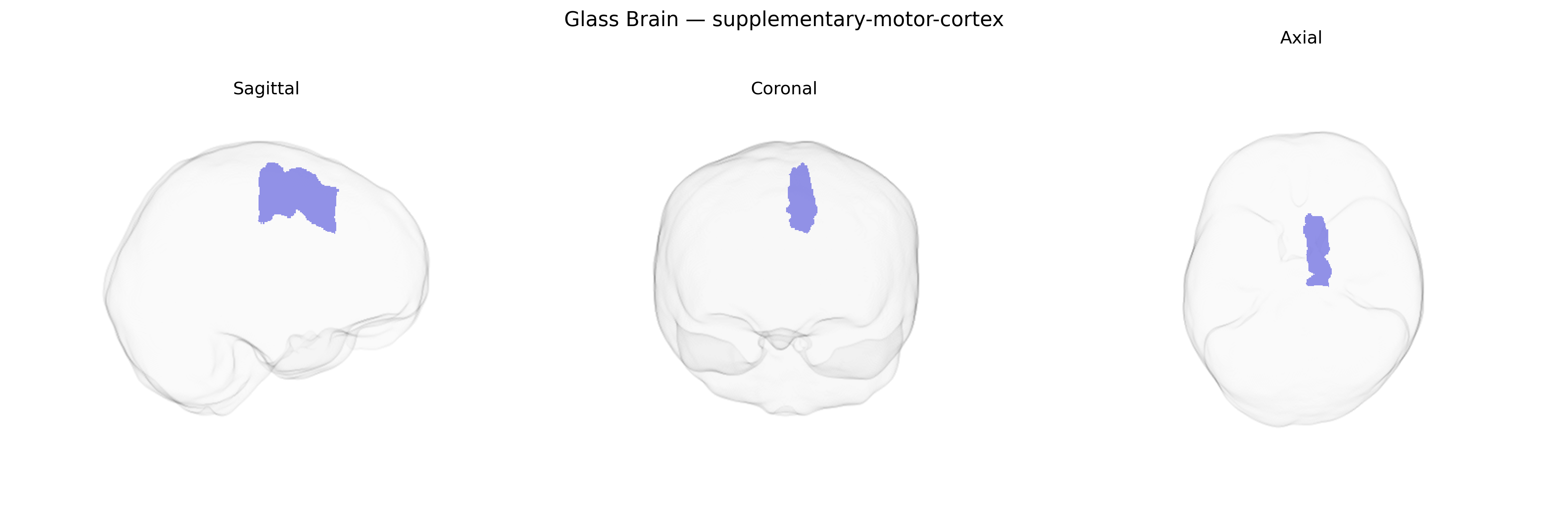
The Left supplementary-motor-cortex, typically encompassing the left supplementary motor area (SMA) on the medial surface of the superior frontal gyrus, is a higher-order motor region involved in planning, initiation, and temporal sequencing of voluntary movements, particularly those that are internally generated, complex, or bimanual. It contributes to motor preparation, coordination between hemispheres, and the transformation of intention into action, integrating information from prefrontal and premotor regions and projecting to primary motor cortex and spinal motor pathways. Functionally, it plays a key role in motor imagery, speech initiation, and the suppression or facilitation of competing motor programs. Lesions in this region can lead to deficits such as impaired initiation of movements (including speech), difficulty with learned motor sequences, and transient akinesia, often with preserved basic motor strength. There is no direct Wikipedia article for “Left supplementary-motor-cortex”; a related entry is Supplementary motor area.
The left supplementary motor cortex (SMA; brainCOLOR Atlas) has been implicated in several genetic and GWAS findings, primarily through studies of cortical morphology, motor function, and neuropsychiatric disorders. Large-scale imaging genetics consortia (e.g., ENIGMA, UK Biobank analyses) have identified common variants influencing cortical thickness and surface area in medial premotor regions encompassing SMA, including loci near genes such as MAPT on 17q21, PAPPA2, and microtubule- or synapse-related genes, though many associations are regionally broad rather than SMA-specific. Variants affecting SMA structure or activation have been linked to motor-related traits and disorders, including dystonia, Parkinson’s disease, Tourette syndrome, and developmental stuttering, where disease-associated loci (e.g., in GNAL, TOR1A, and genes involved in dopaminergic and glutamatergic signaling) show functional connectivity or expression enrichment in SMA circuits. In neuropsychiatry, GWAS of obsessive–compulsive disorder, attention-deficit/hyperactivity disorder, and schizophrenia have highlighted risk loci in genes regulating synaptic plasticity and cortical development (such as CACNA1C, GRIN2A/B, and other glutamatergic and calcium-channel genes) whose polygenic risk scores correlate with altered SMA activation or morphology in task-based and resting-state fMRI. Additionally, genetic variation in FOXP2 and related language-network genes has been associated with altered SMA involvement in speech initiation and sequencing, while motor learning and response-inhibition GWAS point to SMA as a convergent hub for distributed genetic effects on cortical motor control, though direct, SMA-exclusive genetic associations remain relatively sparse and typically emerge as part of broader frontal or medial motor networks.
Overview generated by GPT-4o (2026).
Region ID: 107
Hemisphere: Left
Atlas: brainCOLOR
Full Quality Version: Download MP4
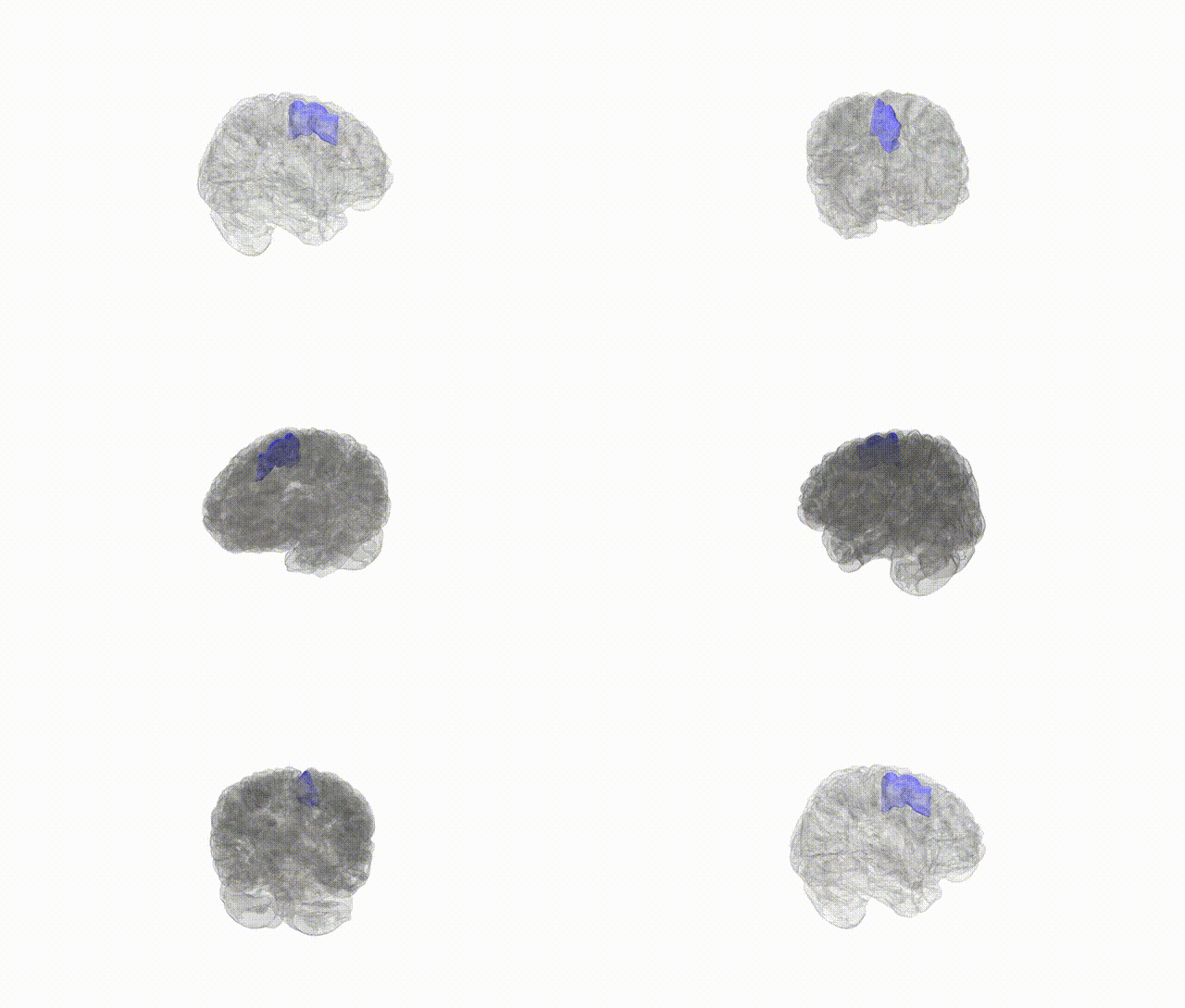
Full Quality Version: Download MP4
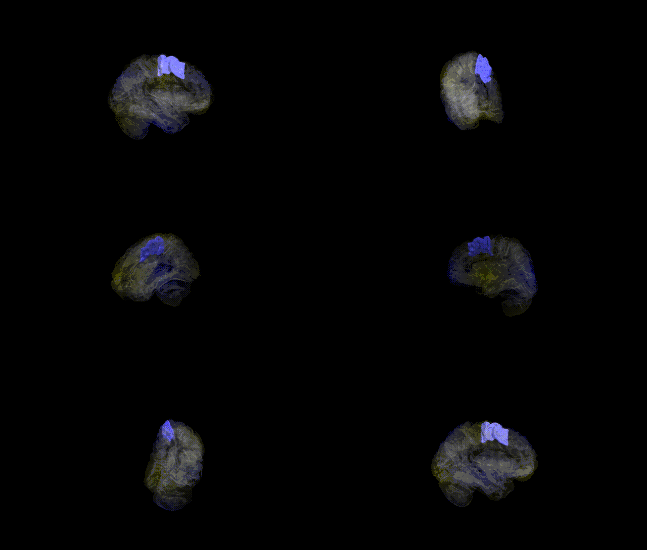
Full Quality Version: Download MP4
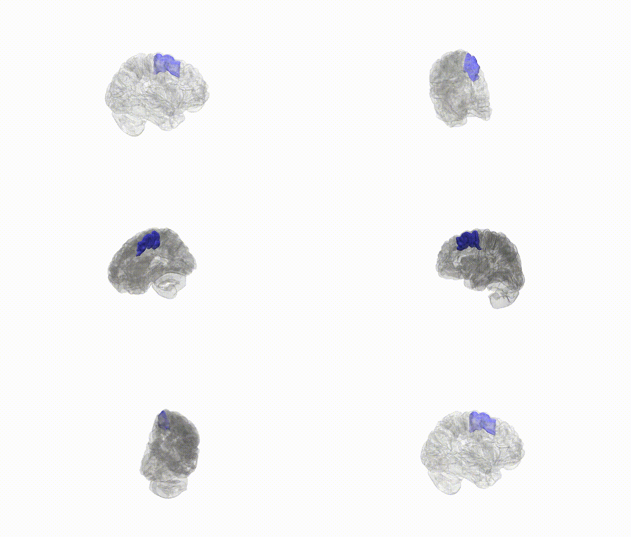
Full Quality Version: Download MP4
Wali Sidiqyar*, Gaurav Rudravaram*, Elyssa M. McMaster, Trent M. Schwartz, Adam M. Saunders, Kurt G. Schilling, Bennett A. Landman "Introducing SPINS: A Shared Public Visualization Library of Neuroanatomical Structures." Medical Imaging with Deep Learning- short paper
This resource is licensed under CC0 1.0 Universal (Public Domain).