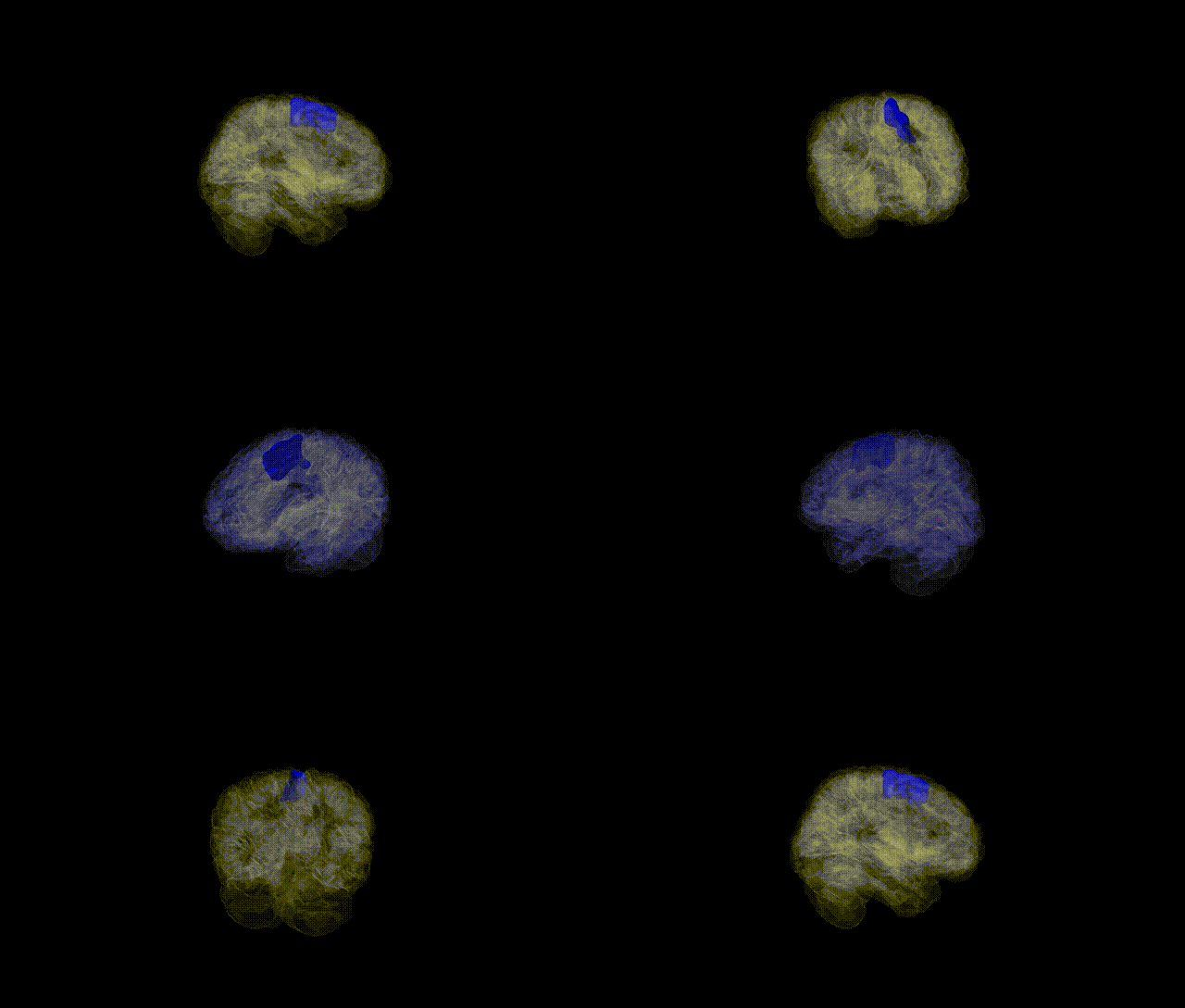
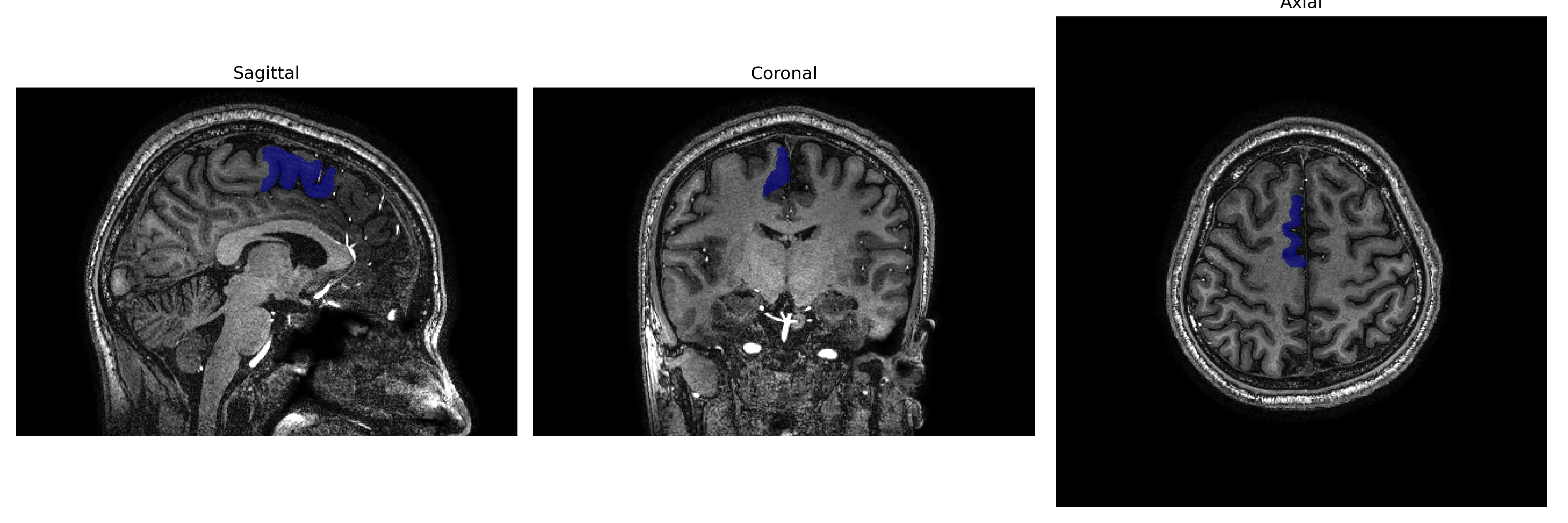
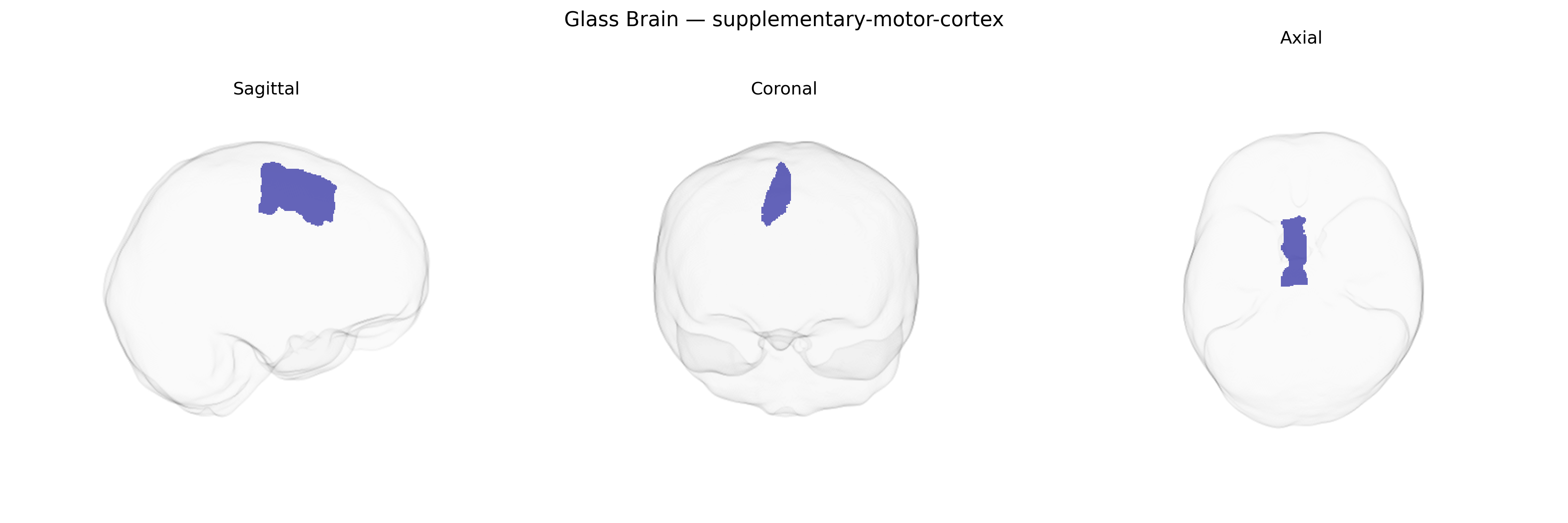
The Right Supplementary Motor Cortex is a medial frontal lobe region located on the dorsal surface of the right hemisphere, anterior to the primary motor cortex and within the medial aspect of Brodmann area 6. It is involved in planning, initiating, and coordinating internally generated movements, particularly complex or sequential motor actions, and contributes to motor imagery, bimanual coordination, and the temporal organization of movements. This region integrates information from prefrontal and parietal areas to shape motor plans before execution and has dense connections with primary motor cortex, premotor cortex, basal ganglia, and thalamus. Functionally, it is implicated in motor preparation and the generation of voluntary actions, and lesions can result in deficits such as akinesia, impaired initiation of speech or movement, and difficulties with learned motor sequences. There is no direct link for the right supplementary motor cortex; a related article is Supplementary motor area.
The right supplementary motor cortex (SMC), as defined in parcellations such as the brainCOLOR atlas, has been implicated in several genetic and GWAS findings, largely via imaging-genetics and disorder-focused studies rather than region-specific GWAS alone. Polygenic influences on cortical surface area and thickness from large consortia (e.g., ENIGMA, UK Biobank) implicate widespread loci—such as variants near genes involved in neurodevelopment and synaptic function (e.g., MAPT region, microtubule and axon guidance genes)—that affect medial frontal and premotor territories including supplementary motor areas. Functionally, right SMC is linked to motor planning, response inhibition, and action sequencing, and genetic risk for disorders featuring motor and executive dysfunction often shows structural or functional alterations here, including in Tourette syndrome and chronic tic disorders (with GWAS implicating genes in synaptic and dopaminergic pathways), obsessive–compulsive disorder, and attention-deficit/hyperactivity disorder, where polygenic risk scores correlate with altered activation in medial frontal–SMC circuitry. In Parkinson’s disease and related movement disorders, variants in genes such as LRRK2 and SNCA, though not specific to SMC, contribute to network-level motor circuit abnormalities that include compensatory or dysregulated right supplementary motor activation. In addition, imaging-genetic work on motor learning and response inhibition suggests that common variants in dopaminergic (e.g., DRD2, COMT) and GABAergic/glutamatergic genes modulate right SMC activation during task performance, linking molecular pathways of neuromodulation and excitation–inhibition balance to this region’s role in higher-order motor control.
Overview generated by GPT-4o (2026).
Region ID: 106
Hemisphere: Right
Atlas: brainCOLOR
Full Quality Version: Download MP4
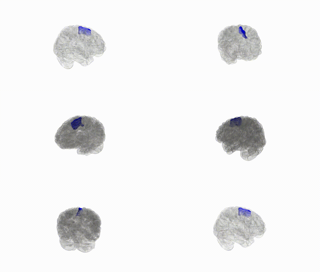
Full Quality Version: Download MP4
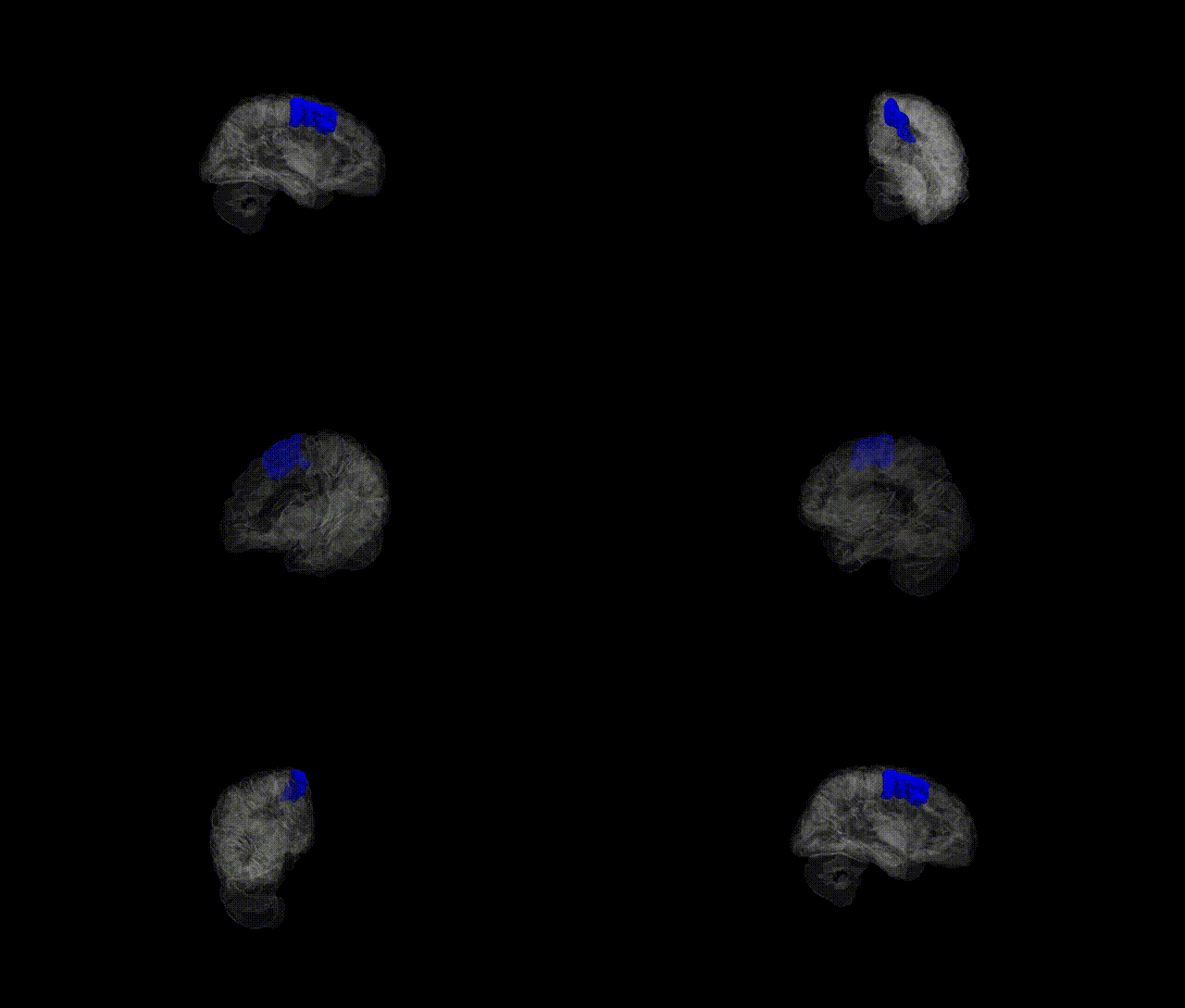
Full Quality Version: Download MP4
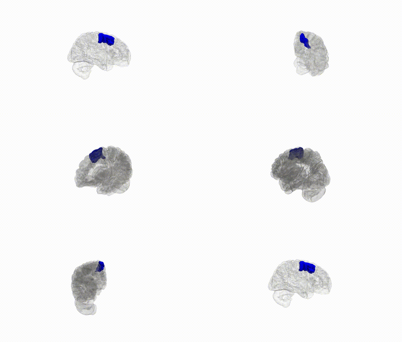
Full Quality Version: Download MP4
Wali Sidiqyar*, Gaurav Rudravaram*, Elyssa M. McMaster, Trent M. Schwartz, Adam M. Saunders, Kurt G. Schilling, Bennett A. Landman "Introducing SPINS: A Shared Public Visualization Library of Neuroanatomical Structures." Medical Imaging with Deep Learning- short paper
This resource is licensed under CC0 1.0 Universal (Public Domain).