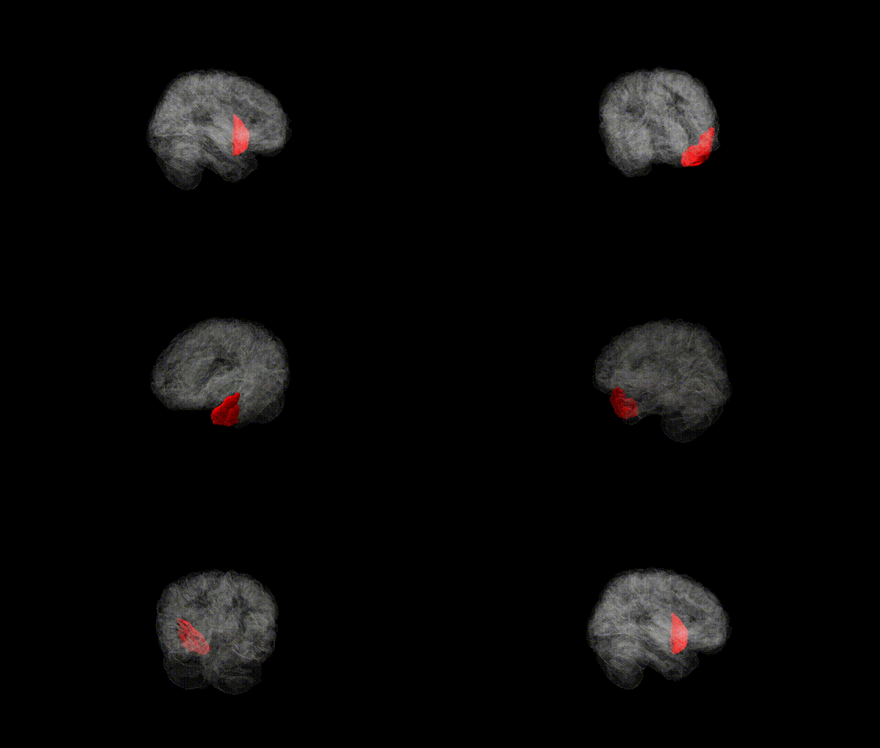
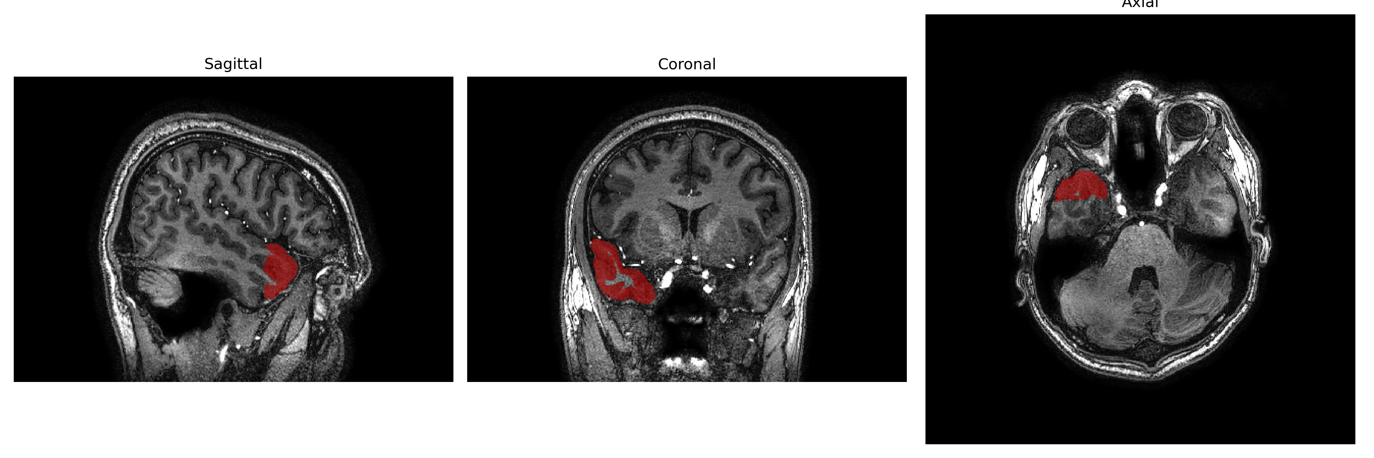
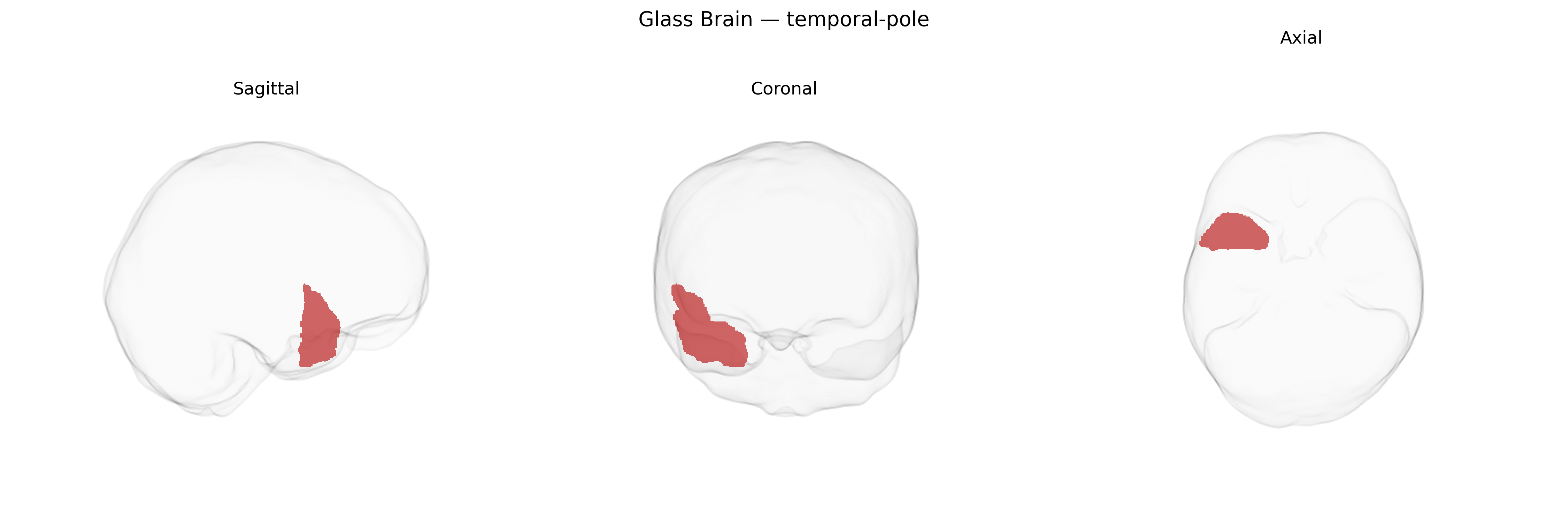
The Right temporal-pole, corresponding roughly to the most anterior portion of the right temporal lobe (Brodmann area 38), is a multimodal associative region integrating high-level auditory, visual, and limbic information. It contributes to social and emotional processing, autobiographical and semantic memory, and the representation of person-specific knowledge, including faces and names. The temporal pole is heavily interconnected with the amygdala, orbitofrontal cortex, and other limbic and paralimbic structures, supporting functions related to emotional valence, theory of mind, and complex language semantics. Lesions or neurodegeneration in this region, such as in semantic variant primary progressive aphasia or frontotemporal lobar degeneration, can lead to profound impairments in conceptual knowledge and social-emotional behavior. There is no direct link for “Right temporal-pole”; see the related structure Temporal pole.
Genetic associations specific to the Right temporal-pole region (as defined in the brainCOLOR Atlas) are not yet well characterized at the single-region level, but several lines of evidence from imaging genetics and GWAS implicate this area in broader temporal and limbic networks. Large-scale brain MRI GWAS (e.g., ENIGMA, UK Biobank–based studies) have identified common variants influencing cortical thickness and surface area in anterior temporal regions, including loci near genes involved in neurodevelopment (such as those affecting synaptic formation, axon guidance, and neuronal migration), though findings are typically reported for aggregated temporal or temporal-pole measures rather than explicitly lateralized right-hemisphere labels. Polygenic risk for schizophrenia, bipolar disorder, major depression, and autism spectrum disorder has been associated with structural and functional alterations in the anterior temporal cortex, with temporal-pole atrophy or dysconnectivity repeatedly noted in frontotemporal dementia (particularly MAPT and GRN mutation carriers) and semantic variant primary progressive aphasia. Additionally, APOE-related risk for Alzheimer’s disease and variants in genes affecting tau and TDP-43 pathology have been linked to patterns of temporal-pole involvement as part of broader temporal-limbic degeneration. Overall, current genetic evidence implicates the Right temporal pole as part of distributed networks affected by neuropsychiatric and neurodegenerative risk variants, but direct, region-specific GWAS findings framed strictly in brainCOLOR Atlas terms remain sparse and usually subsumed under more general anterior or temporal-pole cortical measures.
Overview generated by GPT-4o (2026).
Region ID: 116
Hemisphere: Right
Atlas: brainCOLOR
Full Quality Version: Download MP4
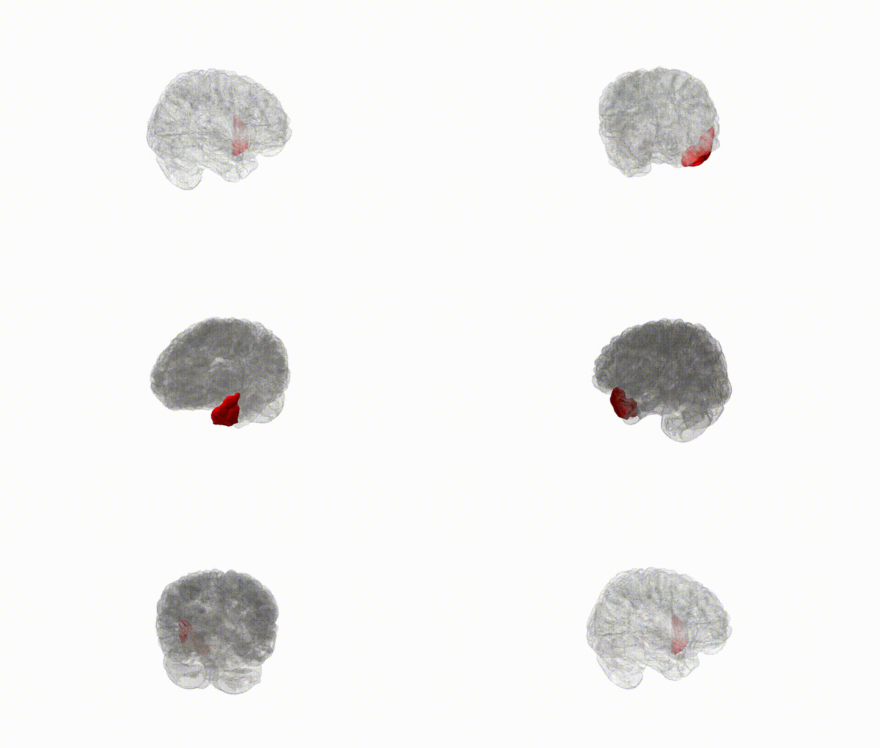
Full Quality Version: Download MP4
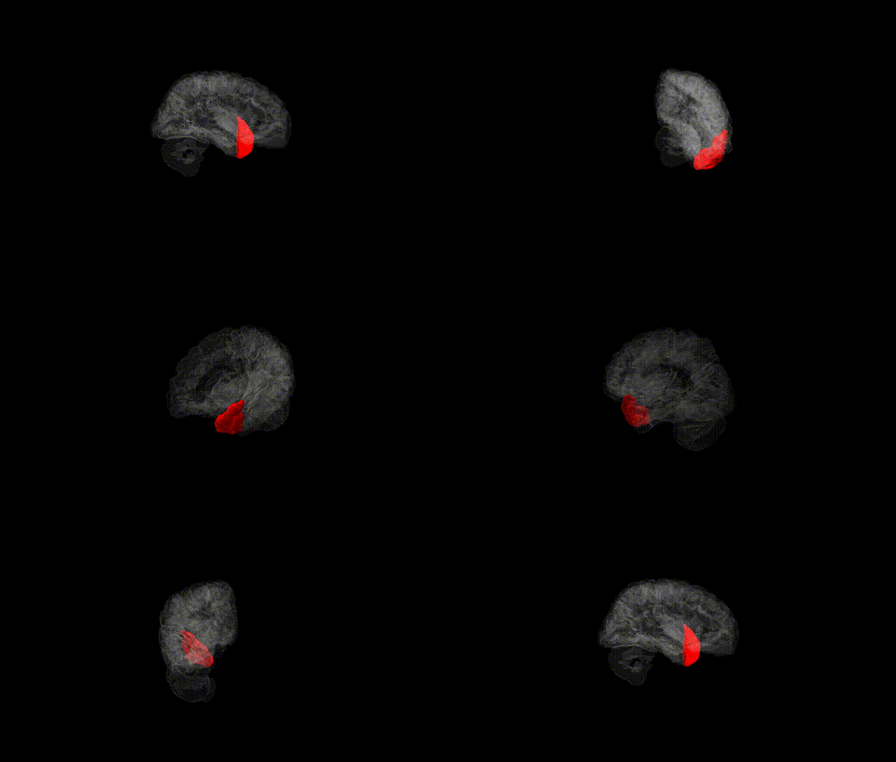
Full Quality Version: Download MP4
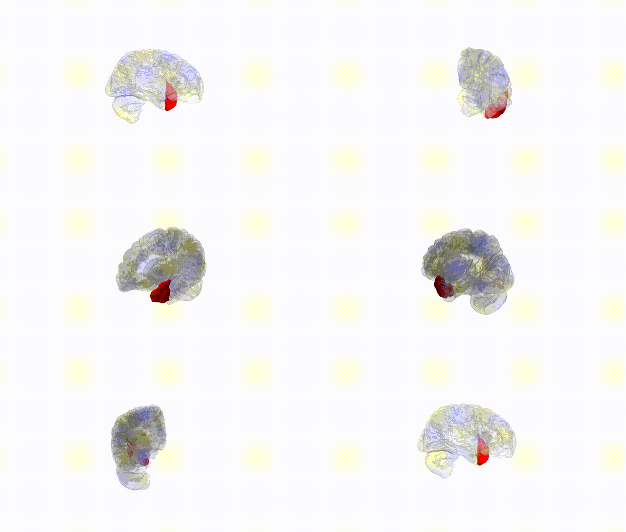
Full Quality Version: Download MP4
Wali Sidiqyar*, Gaurav Rudravaram*, Elyssa M. McMaster, Trent M. Schwartz, Adam M. Saunders, Kurt G. Schilling, Bennett A. Landman "Introducing SPINS: A Shared Public Visualization Library of Neuroanatomical Structures." Medical Imaging with Deep Learning- short paper
This resource is licensed under CC0 1.0 Universal (Public Domain).