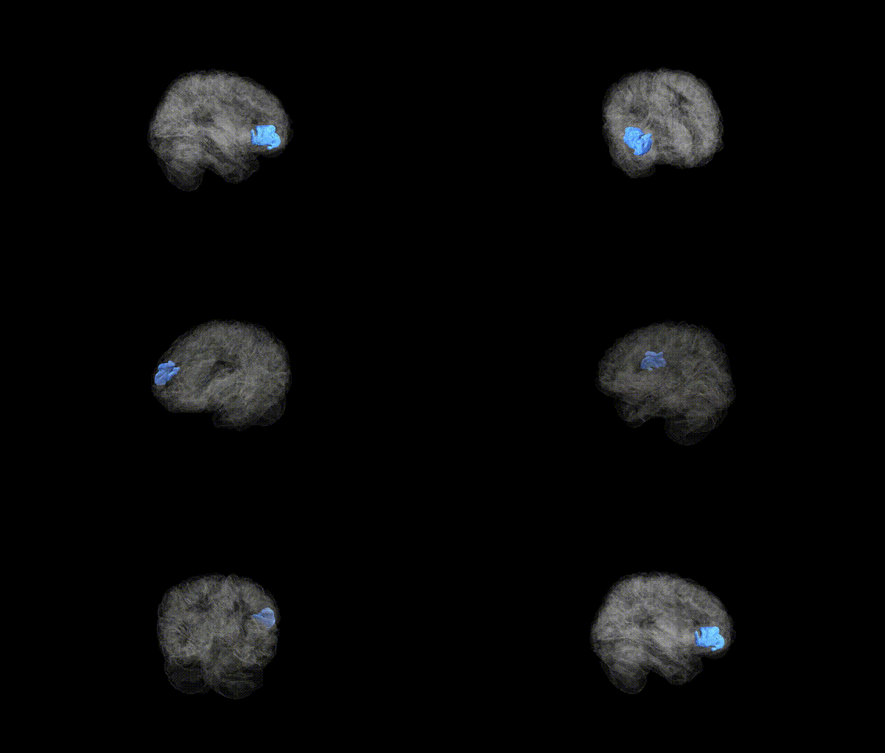
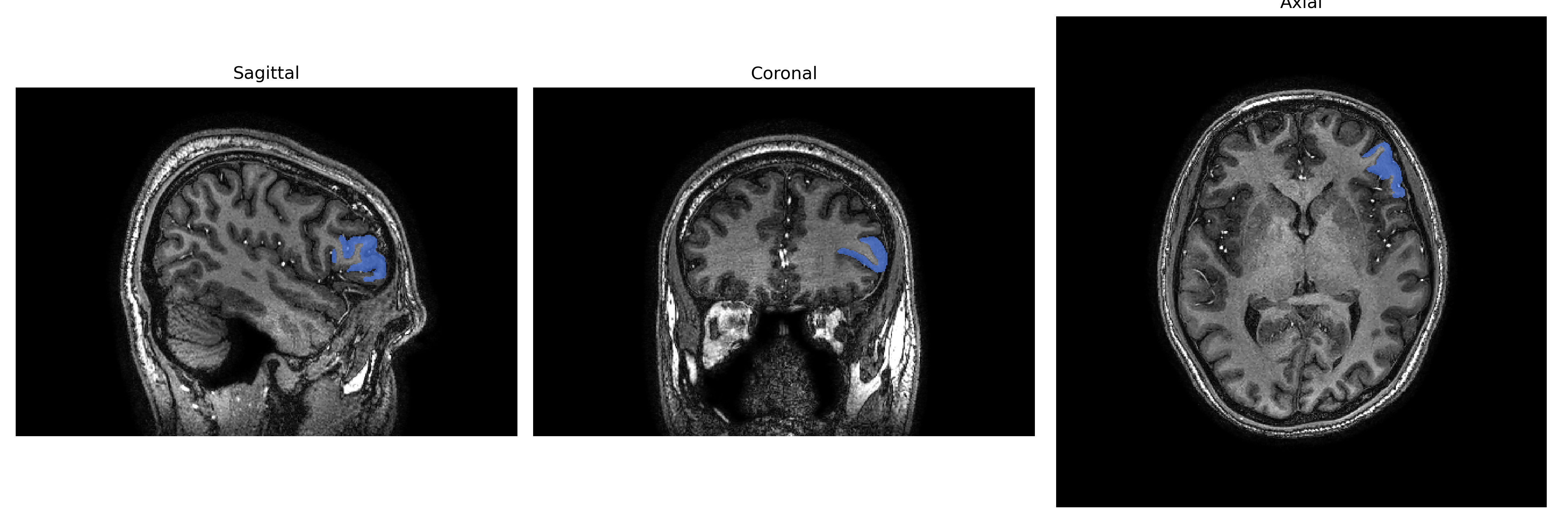
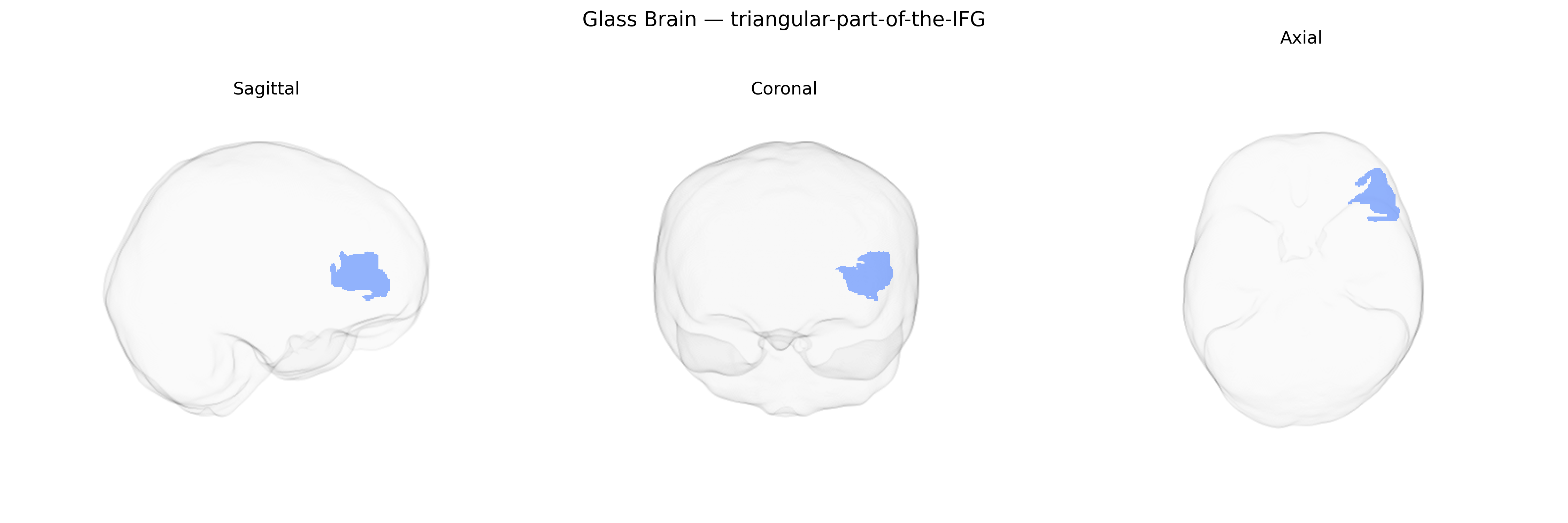
The left triangular part of the inferior frontal gyrus (left triangular IFG) is a cortical subdivision of the inferior frontal gyrus located in the frontal lobe, corresponding largely to Brodmann area 45. It is situated between the pars opercularis posteriorly and the pars orbitalis anteriorly, bordered superiorly by the inferior frontal sulcus and inferiorly by the lateral sulcus (Sylvian fissure). Cytoarchitectonically, it is characterized by granular cortex with distinct layer III and V pyramidal cells and strong connectivity with temporal and parietal association areas, premotor regions, and subcortical structures. Functionally, the left triangular IFG plays a central role in language processing, including semantic retrieval, controlled lexical access, and aspects of syntactic processing, and is often considered part of the broader “Broca’s region.” It is also implicated in cognitive control processes such as response selection, inhibition, and working memory for verbal material. Inferior frontal gyrus
The left triangular part of the inferior frontal gyrus (IFG)—corresponding closely to the anterior/triangular division of Broca’s region—has been implicated in several large-scale imaging genetics and GWAS efforts, although typically under broader labels such as “left inferior frontal gyrus,” “left pars triangularis,” or language-related frontal cortex in atlases including brainCOLOR-based parcellations. Variants near or within genes involved in synaptic development, axon guidance, and cortical patterning (for example, FOXP2, CNTNAP2, KIAA0319, DCDC2, and ROBO1 in dyslexia and language GWAS; as well as more general neurodevelopmental genes such as MAPT, BDNF, and genes in glutamatergic and GABAergic signaling pathways) have been associated with structural or functional measures in this region, including grey matter volume, cortical thickness, and activation during language tasks. GWAS of cortical morphology (e.g., ENIGMA and UK Biobank–based studies) report that common variants across multiple loci influence the size and shape of left inferior frontal subdivisions, with polygenic scores for educational attainment, general cognitive ability, and language-related skills correlating with IFG measures. Moreover, risk variants for neurodevelopmental and psychiatric disorders that affect language and cognitive control—such as autism spectrum disorder, schizophrenia, and attention-deficit/hyperactivity disorder—have been linked to altered structure or activation of the left IFG, suggesting that genetic liability for these conditions partly manifests as differences in this region’s development and function, though specific locus-to-region mappings remain probabilistic and highly polygenic.
Overview generated by GPT-4o (2026).
Region ID: 119
Hemisphere: Left
Atlas: brainCOLOR
Full Quality Version: Download MP4
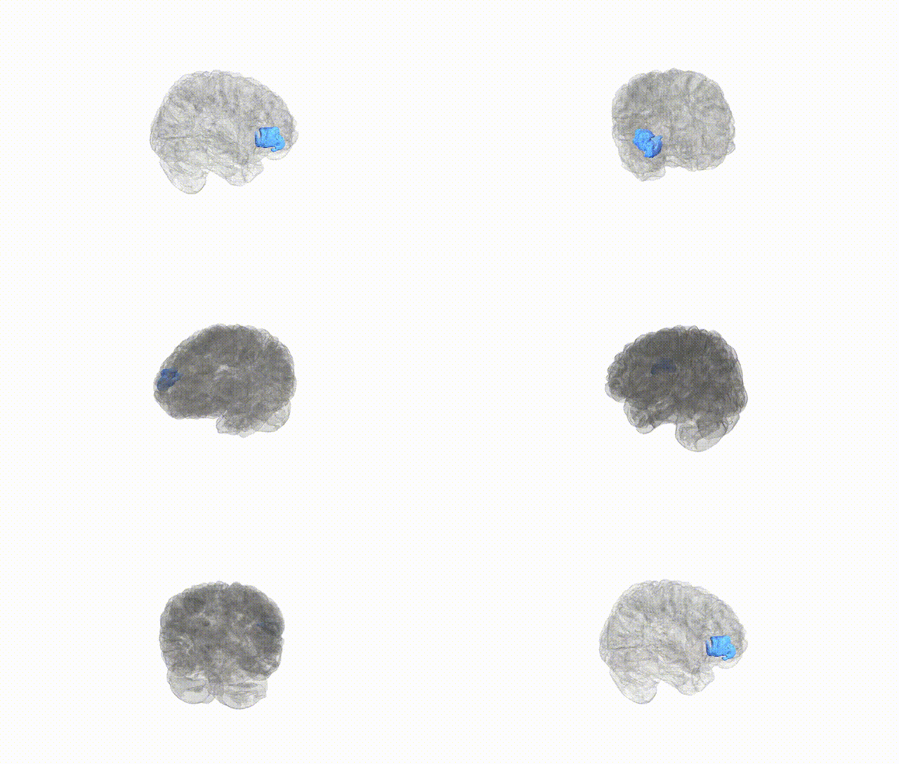
Full Quality Version: Download MP4
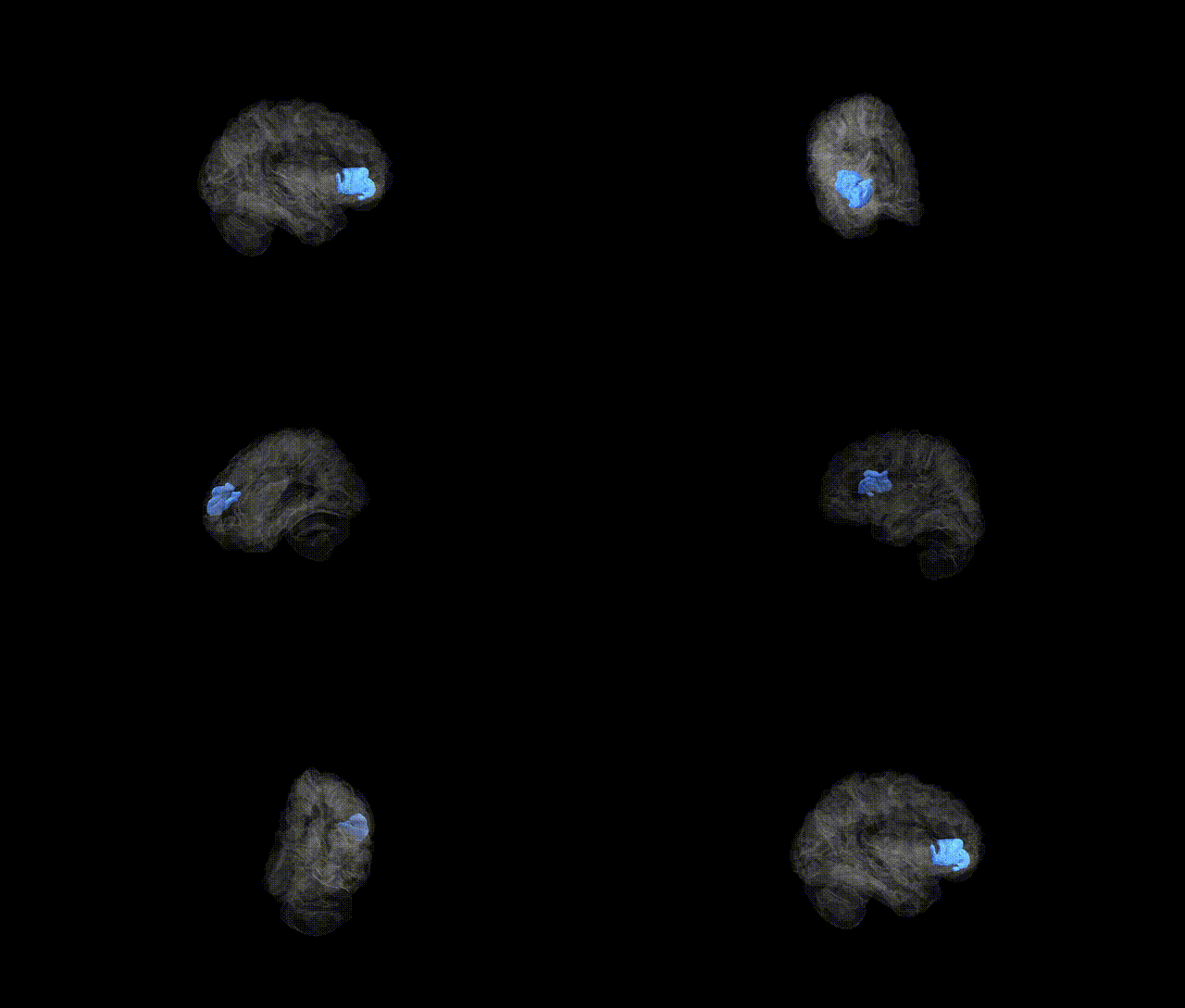
Full Quality Version: Download MP4
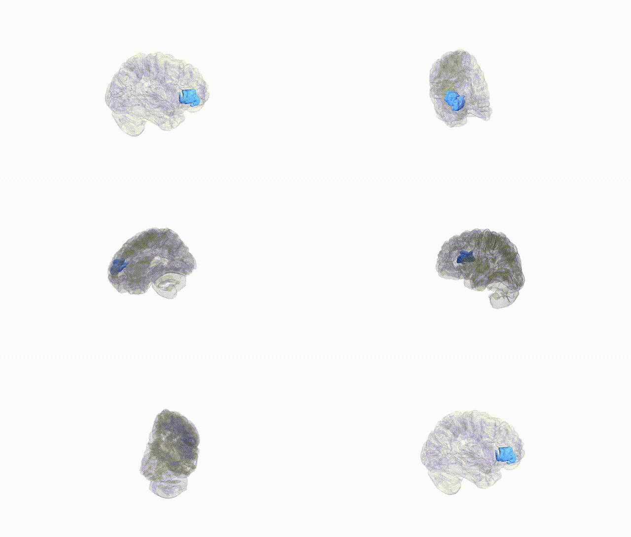
Full Quality Version: Download MP4
Wali Sidiqyar*, Gaurav Rudravaram*, Elyssa M. McMaster, Trent M. Schwartz, Adam M. Saunders, Kurt G. Schilling, Bennett A. Landman "Introducing SPINS: A Shared Public Visualization Library of Neuroanatomical Structures." Medical Imaging with Deep Learning- short paper
This resource is licensed under CC0 1.0 Universal (Public Domain).